Log in    RU  


Main page :: Forum :: Ultrasound cases - Sonograms, cine-loops etc. ::

Torticollis / neoplasia


Tags: Soft tissues sonography, Musculoskeletal sonography, GE Logiq E, Clinical report, Pediatric

   Posts
 
  Torticollis / neoplasia
#1

    Child 1 month old according to the words of the accompanying person, he was examined at two research institutes for a mass on the lateral surface of the neck, in one of the research institutes a biopsy was taken with a suspicious result for a neoplasm, in the other research institutes specialists came to the conclusion that it was torticollis.

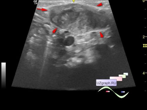
    On the current follow-up ultrasound in the public clinic in the projection of the mass visible to the naked eye in the projection of the left lateral parts of the neck, the ultrasound visualizes a heterogeneous lesion, approximately up to 38 x 13 x 29 mm in size, located in the projection and continuing into the area of normal muscle tissue of the sternocleidomastoid muscle (diff .diagnosis: torticollis, neoplasia, etc.). It is not possible to reliably assess the blood flow at CFM (the child screams, which creates interference).


    external link


 Random 
Ultrasound cases - Pregnancy and urolithiasis
    

Ultrasound cases - Cranial mass
    

Scientific - Playlist Neurosonography

Ultrasound cases - Testicular microlithiasis (microcalcification)


Ultrasound cases - Hygromes of wrist
    

 Dictionary 
BPD

Doppler angle

EDV

Freeze

GA

PA

BSA

MC

IVC

Anatomical M-mode


 Calculators 
Teicholz systolic cardiac function

Myocardial mass

Thyroid volume

Qp:Qs

TI-RADS


 Similar topics 
Ultrasound cases - Penetrating injury of the femur
    

Ultrasound cases - Cephalohematoma
    

Ultrasound cases - Urachus fistula
    

Ultrasound cases - Cranial mass
    

Ultrasound cases - Pilonidal coccyx cyst
    

Ultrasound cases - Palm fistula, in search of pencil lead
    

Ultrasound cases - Postoperative abdominal wall fistula
    

Ultrasound cases - Nucks cyst
    

Ultrasound cases - Paraproctitis
    

Ultrasound cases - Submandibular area lesion