A child 1 year half year ago was sent for soft tissue ultrasound of the soft tissue thickening detected by the parents in the posterior sections of the left shoulder with a transition to the neck, according to ultrasound, a thickening of the subcutaneous soft tissues in this area was detected with a differential diagnosis: lipomatosis, etc.
After the surgeon's consultation, another lesion was revealed below, in the projection of the scapula, after which the child was sent for an X-ray and ultrasound scan of the scapula with suspected exostosis. According to the accompanying person, nothing was found on the X-ray.
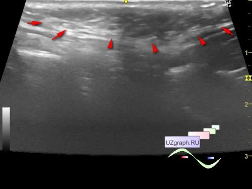
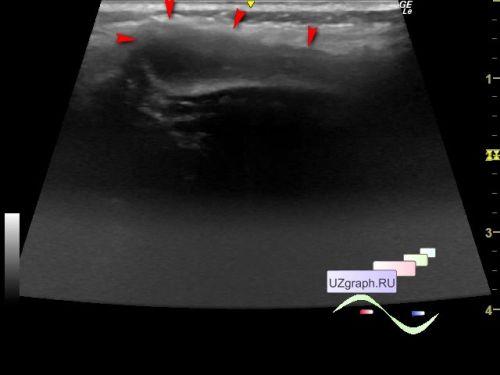
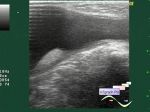
On the current ultrasound in the projection of the lesion visible to the naked eye in the upper part of the left half of the back, with the transition to the neck and shoulder, the ultrasound shows a thickening of the subcutaneous fat compared to the contralateral side (diff. diagnosis: lipomatosis, etc.).
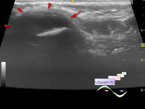
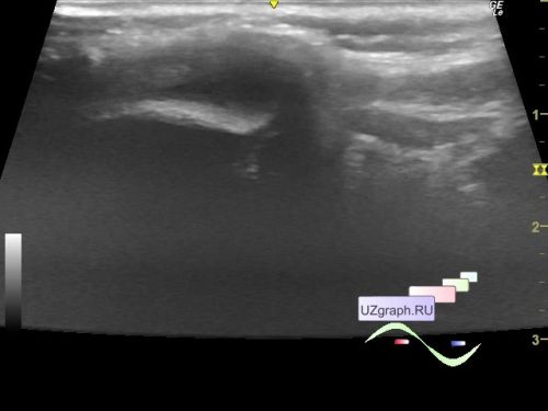
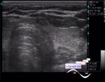
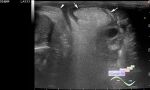
Below this lesion, in the projection of a lesion also visible to the naked eye in the projection of the left scapula, ultrasound visualizes a hypoechoic lesion emanating from the bone of the scapula, without reliable signs of blood flow on the CFM (CFM visualization is difficult due to the go off scale of the CFM scale when the child is crying), up to 32 x 11 x 9 mm (differential diagnosis: exostosis, periostitis, osteomyelitis, lymphoma, myositis, synovitis/ bursitis, etc.)
Consultations of a surgeon, oncologist and MRI of the left scapula were recommended.