Log in    RU  


Main page :: Forum :: Ultrasound cases - Sonograms, cine-loops etc. ::

Hypoechoic spleen lesion


Tags: Abdomen sonography, GE Logiq E, Clinical report, Pediatric

   Posts
 
  Hypoechoic spleen lesion
#1

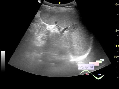
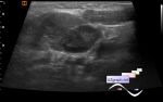
    An 11-year-old teenager came for a second opinion ultrasound after a spleen mass was detected on an abdominal ultrasound because of complaints of abdominal pain.

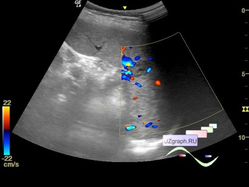
    On ultrasound, the spleen is slightly enlarged, in the middle third of the spleen, a predominantly hypoechoic inclusion with an uneven contour with an anechoic rounded component in the center, at CFM the blood flow is predominantly along the contour of the lesion, up to 18 x 12 x 13 mm in size is visualized.

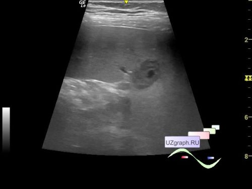
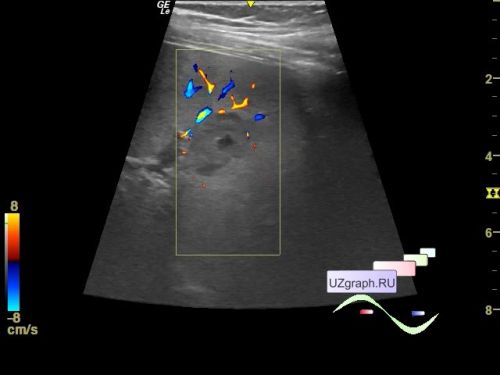
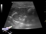
    There is also mesadenitis, thickening of the stomach wall, the appendix is not enlarged.


 Random 
Ultrasound cases - Uterine myoma(fibroid)
    

Ultrasound cases - Pelvic dystopia of the hypoplastic kidney
    

Ultrasound cases - Onion rings sign
    

Ultrasound cases - Lesion type of torsion of Morgagni's hydatide
    

Ultrasound cases - Pain in the right iliac region
    

 Dictionary 
AC

PASP

LMP

TAMEAN

B-mode

TAV

MSD

DPD

EDD

RVOT


 Calculators 
Thyroid volume

Estimated fetal weight by Ultrasound

Qp:Qs

TI-RADS

Body surface area


 Similar topics 
Ultrasound cases - Hirschsprung disease on ultrasound
    

Ultrasound cases - Intestinal Infiltrate
    

Ultrasound cases - Abdominal cryptorchidism
    

Ultrasound cases - Diffuse heterogeneous liver
    

Ultrasound cases - Postoperative abdominal wall fistula
    

Ultrasound cases - Stump appendicitis
    

Ultrasound cases - Liver scar after resection
    

Ultrasound cases - Subdiaphragmatic movable fluid collection (UFO)
    

Ultrasound cases - Lesion of the spleen or the case when it is easy to not notice
    

Ultrasound cases - I’m normal but others ...