Log in    RU  


Main page :: Forum :: Ultrasound cases - Sonograms, cine-loops etc. ::

Good vs Evil - thyroid 4


Tags: Thyroid gland sonography, GE Vivid 3, Images, Video, Clinical report, Adult

   Posts
 
  Good vs Evil - thyroid 4
#1

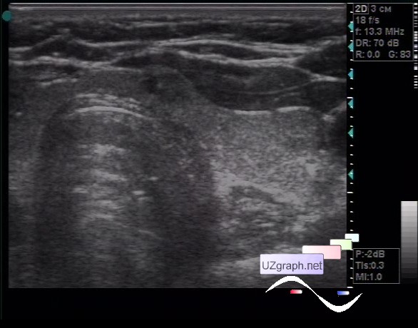
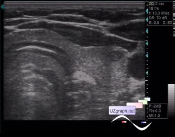
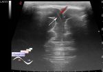
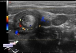
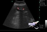
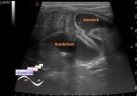
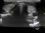
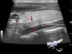
    The woman of 59 years old, has come on a control ultrasound of a thyroid gland concerning nodes.

    At the border of the left lobe and isthmus, a hypoechoic node up to 5x3 mm with hyperechoic microinclusions and with a quaint shape (close to an oval) is visualized, at CFM single signal was visualized (not visible on video) near the node from the side of left lobe; the second node of a similar echostructure, but with a regular oval shape, measuring up to 4x3 mm is visualized in the lateral parts of the left lobe next to the common carotid artery, at CFM without blood flow.

    A puncture is recommended, taking into account the location of one of the nodes in the isthmus region.

    external link

:: attachments(4) ::

:: file 1 ::

:: file 2 ::

:: file 3 ::

:: file 4 ::


 Random 
Ultrasound cases - Birads 4-5
    

Ultrasound cases - Neck lymphangioma
    

Ultrasound cases - Tirads 5
    

Ultrasound cases - Bicuspid aortic valve
    

Ultrasound cases - Torticollis - infiltrate
    

 Dictionary 
B-mode

IVSd

CO

FNA

PSV

MSD

EDD

TVUS

Aliasing doppler artifact

SV


 Calculators 
Type of an infant hip joint

Qp:Qs

Myocardial mass

Gestational age by Ultrasound

Body surface area


 Similar topics 
Ultrasound cases - Tirads 5 multinodular
    

Ultrasound cases - Pediatric Tirads 5
    

Ultrasound cases - Tirads 5
    

Ultrasound cases - Thyroiditis 2
    

Ultrasound cases - Diffuse-focal thyroid changes, parathyroid glands adenomas
    

Ultrasound cases - Accessory Thyroid Lobe
    

Ultrasound cases - Area with hyperechoic dot inclusions
    

Ultrasound cases - Thyroiditis
    

Ultrasound cases - Hypoplasia of the left thyroid lobe
    

Ultrasound cases - Cystic-solid volume of the thyroid gland