Log in    RU  


Main page :: Forum :: Ultrasound cases - Sonograms, cine-loops etc. ::

Cholelithiasis plus 3


Tags: Abdomen sonography, Thyroid gland sonography, GE Vivid 3, Images, Video, Clinical report, Adult

   Posts
 
  Cholelithiasis plus 3
#1

    The patient 51 years old came on the ultrasound of the abdomen and thyroid gland.

    On ultrasound in the gallbladder a stone up to 2.5 cm is visualized.

    external link

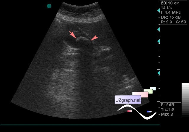
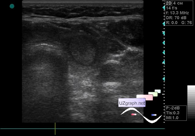
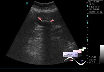
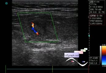
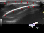
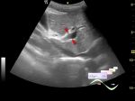
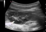
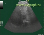
    At ultrasound imaging of the thyroid gland in the left lobe visualizes an iso-echogenic lesion with a hypoechoic rim onto which the blood flow is projected at CFM, up to 1.2 cm in size (adenoma? Other?); as well as the heterogeneity of the right lobe is noted.

    external link

:: attachments(5) ::

:: file 1 ::

:: file 2 ::

:: file 3 ::

:: file 4 ::

:: file 5 ::


 Random 
Ultrasound cases - Periventricular leukomalacia
    

Ultrasound cases - Scrotum hematocele
    

Ultrasound cases - Iliac renal dystopia/ectopia
    

Ultrasound cases - Thyroid lesion
    

Ultrasound cases - Thigh lesion
    

 Dictionary 
Qp-Qs

CO

MSD

TAMEAN

LVESD

Ved

HC

LVEDV

EDV

GS


 Calculators 
Type of an infant hip joint

Volume (ellipse formula)

Myocardial mass

Teicholz systolic cardiac function

Qp:Qs


 Similar topics 
Ultrasound cases - Hirschsprung disease on ultrasound
    

Ultrasound cases - Intestinal Infiltrate
    

Ultrasound cases - Abdominal cryptorchidism
    

Ultrasound cases - Diffuse heterogeneous liver
    

Ultrasound cases - Postoperative abdominal wall fistula
    

Ultrasound cases - Stump appendicitis
    

Ultrasound cases - Liver scar after resection
    

Ultrasound cases - Subdiaphragmatic movable fluid collection (UFO)
    

Ultrasound cases - Lesion of the spleen or the case when it is easy to not notice
    

Ultrasound cases - I’m normal but others ...