Log in    RU  


Main page :: Forum :: Ultrasound cases - Sonograms, cine-loops etc. ::

Non visualization of testicles


Tags: Scrotum sonography, Medison Sonoace R7, Images, Video, Clinical report, Pediatric

   Posts
 
  Non visualization of testicles
#1

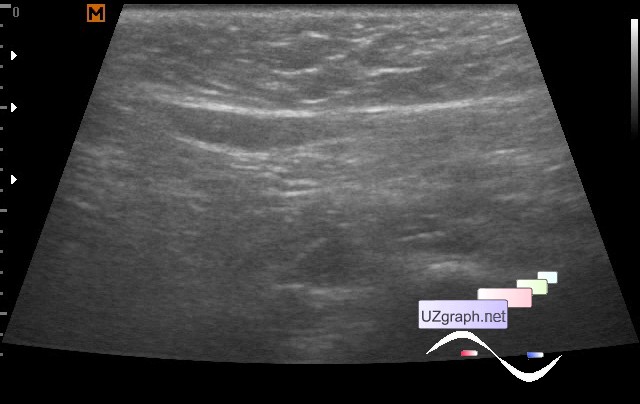
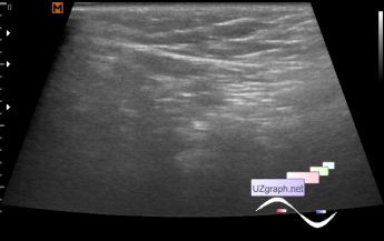
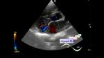
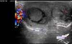
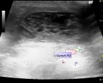
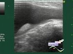
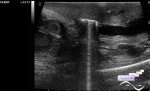
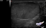
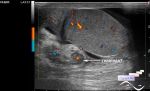
    The child first came to the screening of ultrasound at the age of 4 months, additionally, at the request of the surgeon, the scrotum was examined because the surgeon did not find the testicles. No testicles were found on the ultrasound. Also visually, the penis looked tiny (micropenia?).

    He came back for an examination at the age of 6 months, the testicles are also not visualized, in the projection of the inguinal canals, oval areas with predominantly anehogenous contents are visualized: left to 14x2mm, right to 8x2mm (rudiments of spermatic cord / anorchia? hydrocele?).

    According to the management words, the patient had a second opinion ultrasound by more experienced doctor at the main office of the clinic, who found the testicles but said that they're abnormal.

    I did not see the results of this ultrasound and I do not know on what apparatus the patient was revised and not familiar with this "more experienced" doctor, perhaps just the same echo-picture was interpreted differently.

    external link

:: attachments(5) ::

:: file 1 ::

:: file 2 ::

:: file 3 ::

:: file 4 ::

:: file 5 ::

  16:46 04-09-2018
 
#2

    Similar topic:

    https://m.en.uzgraph.ru/forum/...


 Random 
Admin room - Flash end of life

Ultrasound cases - Subhepatic appendicitis with appendicolith
    

Ultrasound cases - Subdural hemorrhage 2
    

Ultrasound cases - Clinically suspected epididymitis
    

Ultrasound cases - Good vs Evil - femur
    

 Ad 


 Dictionary 
CRL

PD

GS

LVEDD

LVLs

Anatomical M-mode

B-mode

TAV

Aliasing doppler artifact

GA


 Calculators 
Game: Sonographer certification

Body surface area

Estimated fetal weight by Ultrasound

Gestational age by Ultrasound

Measuring distance, area, circumference and angle on an image


 Similar topics 
Ultrasound cases - Abdominal cryptorchidism
    

Ultrasound cases - Neonatal testicular torsion
    

Ultrasound cases - Fibrinous epididymitis
    

Ultrasound cases - Scrotum hematocele
    

Ultrasound cases - Varicocele in a child
    

Ultrasound cases - Hydrocele (dropsy of the scrotum) in a child
    

Ultrasound cases - Air in the scrotum, post surgery condition
    

Ultrasound cases - Multi-chamber scrotum cyst
    

Ultrasound cases - Diffuse changes of the testicle in a teenager (orchitis)
    

Ultrasound cases - Thickening of the spermatic cord in a teenager
    

 Ad