Log in    RU  


Main page :: Forum :: Ultrasound cases - Sonograms, cine-loops etc. ::

Thyroid lesions


Tags: Thyroid gland sonography, Medison Sonoace R7, Images, Video, Clinical report, Adult

   Posts
 
  Thyroid lesions
#1

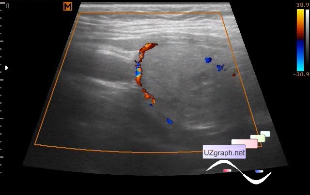
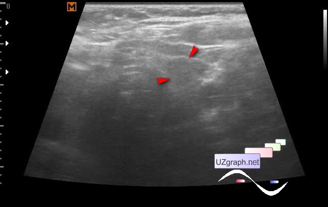
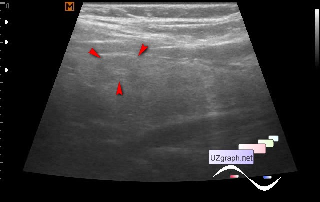
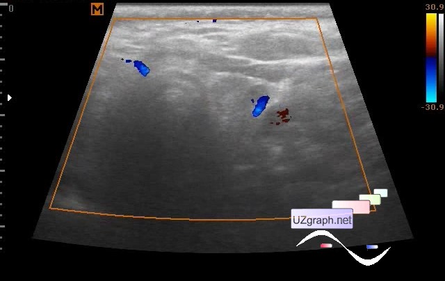
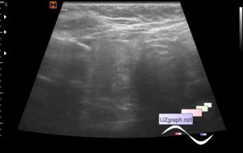
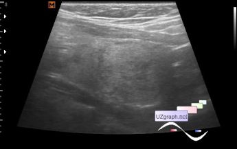
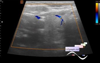
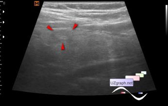
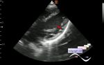
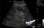
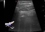
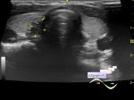
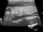
    A woman of 50 years with complaints about the thyroid gland, she tells that it hinders her.

    In the right lobe: predominantly isoechogenic non-uniform lesion of 3x2 cm with a hypoechogenic rim, which is stained at CFM, oval shaped (adenoma? else?); and also single predominantly anechogenous lesion up to 5 mm (colloid node? else?).

    In the left lobe: iso-hypoechoic lesion up to 1 cm, the blood flow along the contour at CFM, and also 3-4 predominantly anechoic lesions, some with hyperechoic microinclusions, up to 5 mm (colloidal nodes, else?).

    external link

:: attachments(9) ::

:: file 1 ::

:: file 2 ::

:: file 3 ::

:: file 4 ::

:: file 5 ::

:: file 6 ::

:: file 7 ::

:: file 8 ::

:: file 9 ::


 Random 
Ultrasound cases - Hernia of the linea alba / lipoma
    

Ultrasound cases - Uterine myoma(fibroid)
    

Ultrasound cases - Cholesterol pseudopolyp?
    

Ultrasound cases - Abdominal syndrome, two in one
    

Ultrasound cases - Inguinal hernia with an ovary
    

 Dictionary 
BPD

HC

LVEDD

RAP

Aliasing doppler artifact

PA

VTI

EDV

EFW

CRL


 Calculators 
Type of an infant hip joint

Body surface area

Thyroid volume

Pulmonary artery pressure

TI-RADS


 Similar topics 
Ultrasound cases - Tirads 5 multinodular
    

Ultrasound cases - Pediatric Tirads 5
    

Ultrasound cases - Tirads 5
    

Ultrasound cases - Thyroiditis 2
    

Ultrasound cases - Diffuse-focal thyroid changes, parathyroid glands adenomas
    

Ultrasound cases - Accessory Thyroid Lobe
    

Ultrasound cases - Area with hyperechoic dot inclusions
    

Ultrasound cases - Thyroiditis
    

Ultrasound cases - Hypoplasia of the left thyroid lobe
    

Ultrasound cases - Cystic-solid volume of the thyroid gland