Log in    RU  


Main page :: Forum :: Ultrasound cases - Sonograms, cine-loops etc. ::

Left & right ventricular non-compaction


Tags: Cardiac sonography(Echocardiography), Medison Sonoace R7, Images, Video, Clinical report, Pediatric

   Posts
 
  Left & right ventricular non-compac...
#1

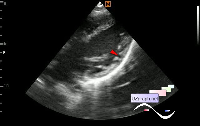
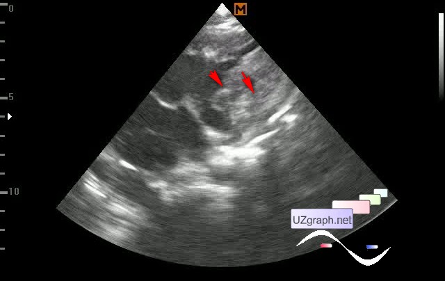
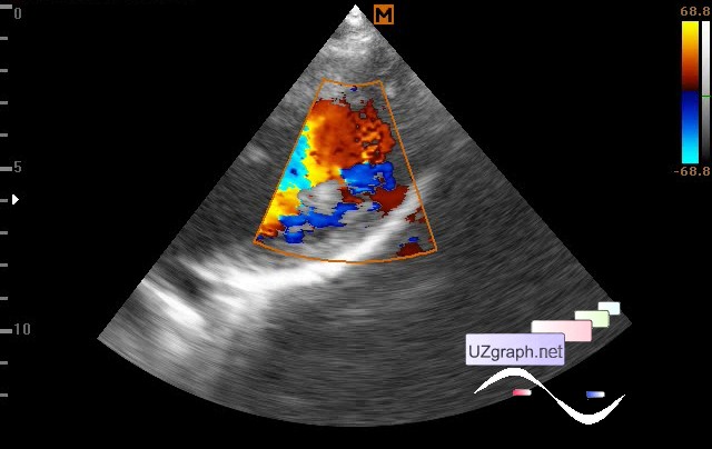
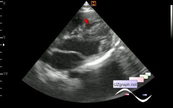
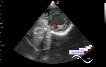
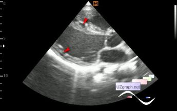
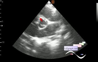
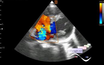
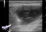
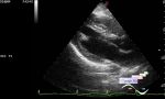
    A child of 10 years old, in the EMR a diagnosis of VSD of the muscular part of 2 mm is indicated.

    According to the accompanying person child was observed in one Moscow Children' s Clinical Hospital with this diagnosis, where they said that it was okay, but they did not recommend sports. Child play sports before.

    At the current ultrasound, the wall of the LV (mainly posterior-lateral) with an uneven internal contour of the type of polyps with the bulbous spaces between, which are stained on the CFM. In the center of IVS anechoic spaces are visualized, presumably communicating with each other and with the RV cavity, in some oblique views dividing IVS into two parts, especially in 2ch positions, creating the appearance of perimembranous VSD or two IVS near (noncompact myocardium?).

    The aortic valve opens as a two-leaf valve.

    At CFM also visualized the regurgitation of 2-3st. at the TV.

    With the red stream at the apex, I still could not decide whether it was a muscular VSD or another area of the noncompact myocardium in the RV.

    external link

:: attachments(10) ::

:: file 1 ::

:: file 2 ::

:: file 3 ::

:: file 4 ::

:: file 5 ::

:: file 6 ::

:: file 7 ::

:: file 8 ::

:: file 9 ::

:: file 10 ::

  11:17 23-04-2020
  apical-basal muscle bundle
#2

    "The apical-basal muscle bundle refers to an accessory papillary muscle or muscle bundle that extends from the LV apex and inserts into the basal myocardium without insertion into the mitral leaflet. This condition is commonly present in patients with HCM (63%) and in gene-positive family members (60% vs 10% of controls)."

    external link

    So its supposed that at the following image - one part is the IVS and the other - the "apical-basal muscle bundle":


 Random 
Ultrasound cases - Pneumonia
    

Ultrasound cases - Hematoma of the gluteal region
    

Ultrasound cases - Good vs Evil
    

Ultrasound cases - Meconium peritonitis and ascites
    

Ultrasound cases - Portal hypertension
    

 Dictionary 
Doppler angle

FL

IVC

TVUS

PD

PASP

EFW

VTI

LN

Gain


 Calculators 
Type of an infant hip joint

Volume (ellipse formula)

Resistive index

TI-RADS

Gestational age by Ultrasound


 Similar topics 
Ultrasound cases - Bicuspid aortic valve 2
    

Ultrasound cases - Mitral valve cyst
    

Ultrasound cases - Electrode in the right heart after installing of heart pacemaker
    

Ultrasound cases - Chylopericardium, lung atelectasis, cardiomegaly, ascites
    

Ultrasound cases - Asymmetric LVH, aortic regurgitation
    

Ultrasound cases - Coronary fistula from LCA?
    

Ultrasound cases - Floating IAS aneurysm
    

Ultrasound cases - Aortic Stenosis
    

Ultrasound cases - Floating Accessory Pulmonary Valve Tissue
    

Ultrasound cases - Hyperechoic linear structure in the left atrium