Log in    RU  


Main page :: Forum :: Ultrasound cases - Sonograms, cine-loops etc. ::

Lower extremity swelling


Tags: Soft tissues sonography, Musculoskeletal sonography, Vascular sonography, Images, Video, Clinical report, Esaote MyLab 70, Pediatric

   Posts
 
  Lower extremity swelling
#1

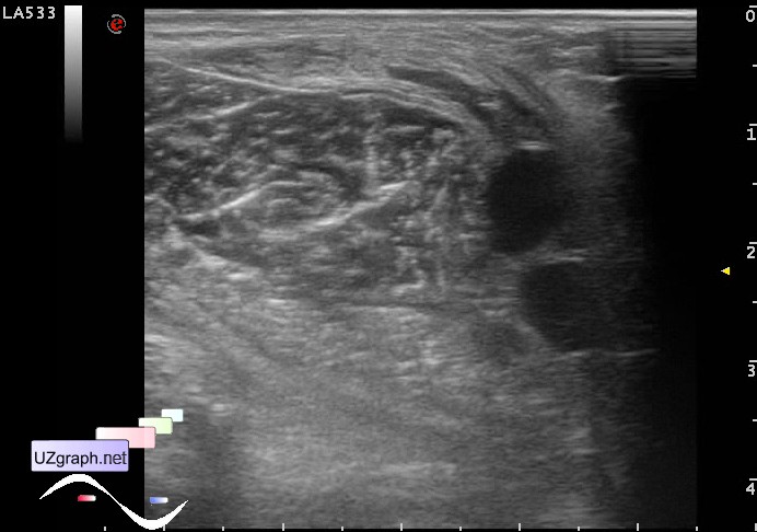
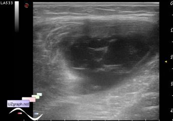
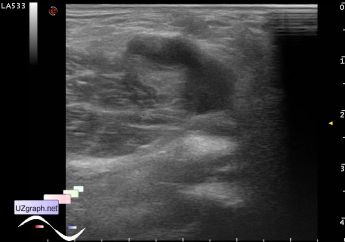
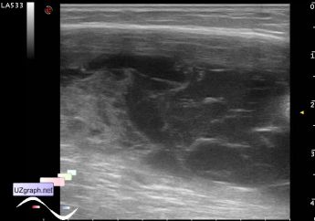
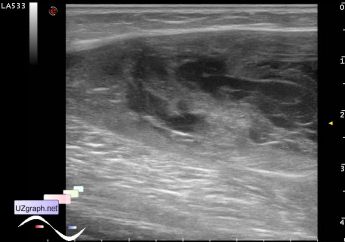
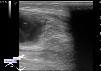
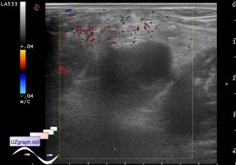
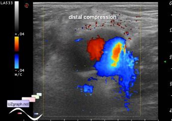
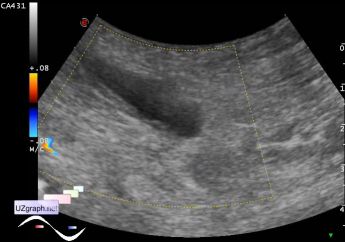
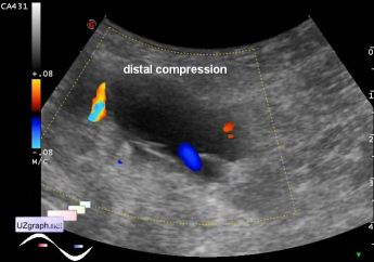
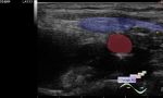
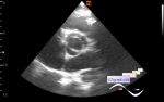
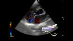
    Teenager with complaints about recently former shin swelling for an unknown reason, aims to ultrasound of lower extremity veins with suspected lower extremity deep vein thrombosis.

    At ultrasound the deep veins are normal, at the medial surface of the popliteal fossa visualized tubular mass looks like a cyst, at CFM without blood flow but during shin compression with flow at CFM(phlebectasia?), at the medial surface of almost full length of the shin large mass with multiple partitions looks like encapsuled fluid (hematoma? teenager denies trauma) without blood flow at CFM. No clear border between this two(?) masses were found.

    Later examined by another sonologist(looked together), at standing position the popliteal fossa mass was colored by CFM, however the doctor made conclusion that this is a Baker' s cyst.

    external link

:: attachments(12) ::

:: file 1 ::

:: file 2 ::

:: file 3 ::

:: file 4 ::

:: file 5 ::

:: file 6 ::

:: file 7 ::

:: file 8 ::

:: file 9 ::

:: file 10 ::

:: file 11 ::

:: file 12 ::


 Random 
Ultrasound cases - Midgut malrotation with volvulus
    

Ultrasound cases - Thickening of the sternocleidomastoid
muscle


Ultrasound cases - Interatrial Communication
    

Ultrasound cases - Bilateral paraproctitis
    

Ultrasound cases - Cyst of spermatic cord
    

 Dictionary 
TAMEAN

GA

EDD

Doppler angle

LVPWd

TI-RADS

EF

LMP

B-mode

TVUS


 Calculators 
Pressure gradient

Type of an infant hip joint

Thyroid volume

Body surface area

Qp:Qs


 Similar topics 
Ultrasound cases - Penetrating injury of the femur
    

Ultrasound cases - Cephalohematoma
    

Ultrasound cases - Urachus fistula
    

Ultrasound cases - Cranial mass
    

Ultrasound cases - Pilonidal coccyx cyst
    

Ultrasound cases - Palm fistula, in search of pencil lead
    

Ultrasound cases - Postoperative abdominal wall fistula
    

Ultrasound cases - Nucks cyst
    

Ultrasound cases - Paraproctitis
    

Ultrasound cases - Submandibular area lesion