Log in    RU  


Main page :: Forum :: Ultrasound cases - Sonograms, cine-loops etc. ::

PO suture


Tags: Soft tissues sonography, GE Vivid 3, Images, Video, Clinical report, Adult

   Posts
 
  PO suture
#1

    The patient 56 years old came to control ultrasound of LNs (axillary, supra - and subclavian) and PO (postoperative) suture.

    From anamnesis it is known that the patient underwent resection of melanoma of the lumbar region, as well as MTS in the axillary LN on the same side. The size of the melanoma was about 2 cm, PO scar visually about 5 cm or more.

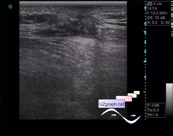
    At ultrasound LN in these areas are not visualized, PO scar is visualized as a hetero-echogenic defect of tissues (dif.diagnosis: granuloma, etc.).

    external link

:: attachments(2) ::

:: file 1 ::

:: file 2 ::


 Random 
Admin room - International day of radiology

Ultrasound cases - Gynecomastia
    

Ultrasound cases - Unconscious, history, clinic, US
    

Ultrasound cases - Palm fistula, in search of pencil lead
    

Ultrasound cases - Nobody knows what it is
    

 Dictionary 
Gain

SF

Qp-Qs

MSD

TAMEAN

FVI

M-mode

PASP

PRF

EDV


 Calculators 
Teicholz systolic cardiac function

Pressure gradient

Thyroid volume

Pulmonary artery pressure

Resistive index


 Similar topics 
Ultrasound cases - Penetrating injury of the femur
    

Ultrasound cases - Cephalohematoma
    

Ultrasound cases - Urachus fistula
    

Ultrasound cases - Cranial mass
    

Ultrasound cases - Pilonidal coccyx cyst
    

Ultrasound cases - Palm fistula, in search of pencil lead
    

Ultrasound cases - Postoperative abdominal wall fistula
    

Ultrasound cases - Nucks cyst
    

Ultrasound cases - Paraproctitis
    

Ultrasound cases - Submandibular area lesion