Log in    RU  


Main page :: Forum :: Ultrasound cases - Sonograms, cine-loops etc. ::

Autosomal recessive polycystic kidney disease (ARPKD)


Tags: Urinary tract sonography, Esaote MyLab 70, Clinical report, Pediatric

   Posts
 
  Autosomal recessive polycystic kidn...
#1

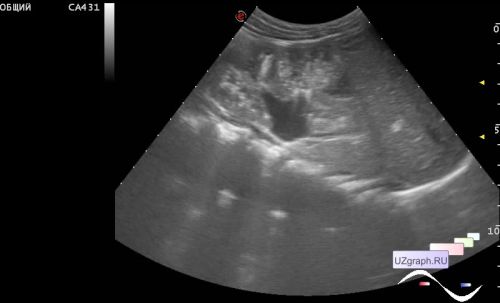
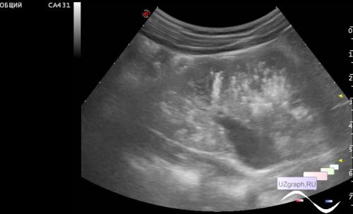
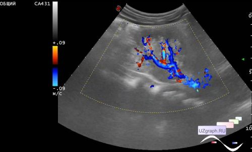
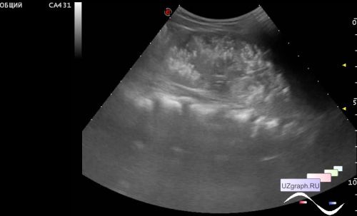
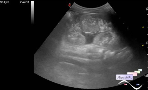
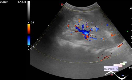
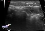
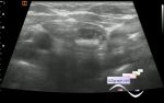
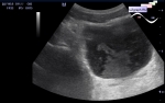
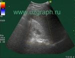
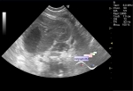
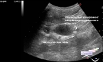
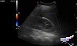
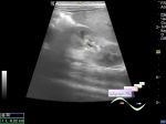
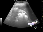
    A 5 years old child with suspected polycystic kidney disease, a follow-up ultrasound was prescribed.

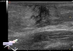
    On ultrasound, the kidneys are enlarged in size, the structure is diffusely changed due to multiple hyperechoic microinclusions, giving a comet tail artifact, in both kidneys.


 Random 
Scientific - Lecture about LUS at COVID-19
    

Ultrasound cases - Uterine cervix hyperechogenicity
    

Ultrasound cases - Ascites & cholelithiasis
    

Ultrasound cases - Clinically suspected appendicitis 3
    

Ultrasound cases - Suspected intestinal pneumatosis
    

 Dictionary 
SV

CFM

GS

HR

TVUS

PG

GA

BSA

PSV

PADP


 Calculators 
Body surface area

Pulmonary artery pressure

Myocardial mass

Teicholz systolic cardiac function

Resistive index


 Similar topics 
Ultrasound cases - Bladder sludge
    

Ultrasound cases - Bright kidneys
    

Ultrasound cases - Ileostoma stenosis and nephropathy
    

Ultrasound cases - Renal pelvis clot
    

Ultrasound cases - Renal pelvis valve-stone
    

Ultrasound cases - Condition after treatment of a kidney cyst
    

Ultrasound cases - Hydronephrotic transformation & polypous bladder wall
    

Ultrasound cases - Pyelitis, cystitis
    

Ultrasound cases - Lesion in the projection of the adrenal gland 2
    

Ultrasound cases - Hyperechoic renal sinus with acoustic shadow