Main page :: Forum :: Ultrasound cases - Sonograms, cine-loops etc. :: Tags: Chest sonography Medison Sonoace R7 Images Video Clinical report Pediatric Moderator :
User:
admin Registered: 23-09-2013
18:24 13-10-2017 #1
A casual find.
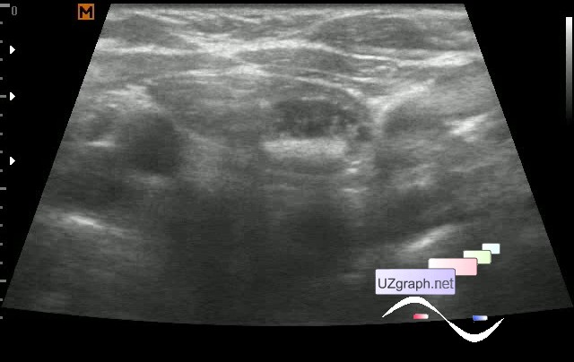
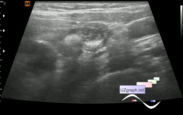
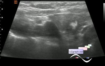
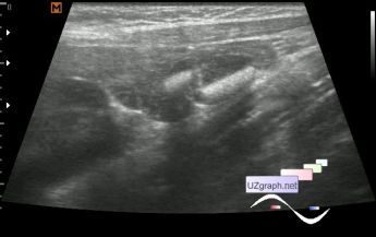
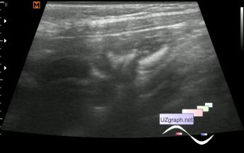
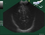
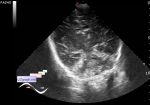
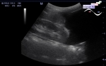
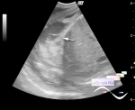
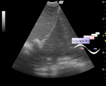
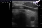
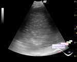
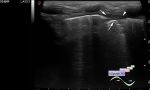
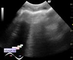
The child of 8 years has come on thyroid US , a thyroid gland without features.
My eye see something in the thymus and here we go ...
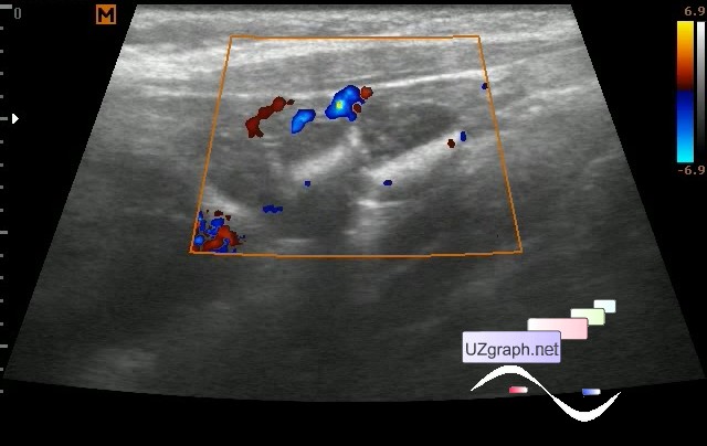
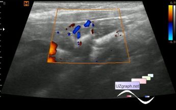
The thymus gland is not enlarged with diffusive-heterogeneous structure due to lesions of different sizes, in the right lobe there are 2 hyperechoic lesions up to 4 mm, in the left lobe there are two intimately located lesions of a mixed hyper / hypoechoic structure, at CFM there is a bloodflow predominantly along the contour, measuring up to 1 cm each.
It was recommended CT of the thorax, the child was sent for consultation to an oncologist in the profile center.
external link
:: file 1 ::
:: file 2 ::
:: file 3 ::
:: file 4 ::
:: file 5 ::
:: file 6 ::
:: file 7 ::
:: file 8 ::
:: file 9 ::
:: file 10 ::
:: file 11 ::
HTML5 video plugin not supported!
Carpe Diem
Right now my main version is a teratoma( teratomas).
Carpe Diem
The child was consulted in the oncological center, where according to the provided papers only ultrasound was performed:
" Clinical diagnosis:
... The pathology of the thymus is not revealed.
...
Ultrasound ... the thymic tissue is moderately diffuse nonuniform, without nodular and cystic lesions. Density parenchymal, uneven.
...
Conclusion:
Signs of tumor lesion and inflammatory changes are not revealed. "
Carpe Diem
Despite the fact that to child in the oncological center was recommended the control ultrasound after 6 months, I called the child to control ...
Lesions has not gone away, only slightly changed the structure, shape, size.
Thanks to my colleague who sent me the link:
external link
Where in the diagnostic algorithm it is clearly prescribed that if there are mass in the thymus other variants than CT or MRI can' t be.
I showed it to the parent, I hope it worked.
And explained that ultrasound carried out even by PhD do not replace CT!
Again recommended CT / MRI. It is planned to send the patient to another hospital.
Carpe Diem
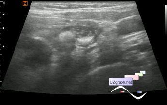
Video - control ultrasound after 2 weeks.
:: file 1 ::
HTML5 video plugin not supported!
Carpe Diem
Several colleagues expressed an opinion that this could be cysts.
I rummaged on the site medison.ru:
external link
But the cysts on their echograms looks like simple cysts, but not on mine.
Here are another sources:
external link
external link
Quote from the last:
" Postnatally, sonography, computed tomography, and magnetic resonance imaging may play a role in diagnosis and identification of the thymic origin.
...
Resection of thymic cysts is optimal for prevention or treatment of complications and because neoplasia cannot be definitively excluded."
Although these sources noted the complete regression of cysts without surgery.
Maybe it' s true, but in my case the echograms do not looks like simple cysts.
Carpe Diem
Mediastinal masses-transthoracic ultrasonography aspects -
external link
And I also did not find anything like in my case...
Carpe Diem