Log in    RU  


Main page :: Forum :: Ultrasound cases - Sonograms, cine-loops etc. ::

Diaphragmatic hernia, condition after surgery


Tags: Chest sonography, GE Logiq Book XP, Clinical report, Pediatric

   Posts
 
  Diaphragmatic hernia, condition aft...
#1

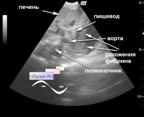
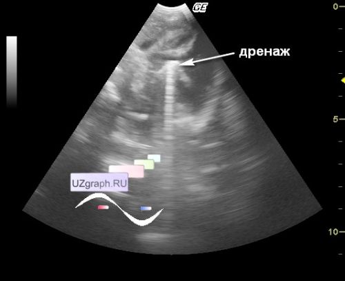
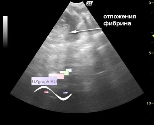
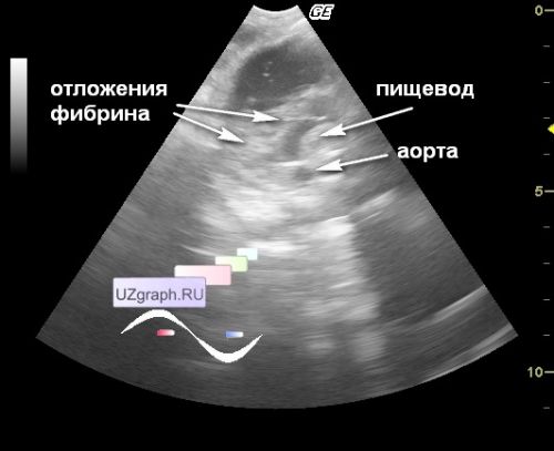
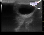
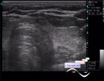
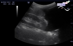
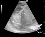
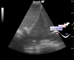
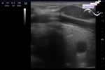
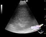
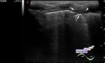
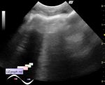
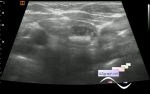
    Newborn in the ICU of the Children's City Clinical Hospital after surgery of left-sided diaphragmatic hernia. An ultrasound of the pleural spaces was prescribed for fluid.

    On ultrasound, the entire left pleural cavity is filled with fluid, the lung is not visible (presumably hypoplastic), a tubular structure with a reverberation artifact (presumably drainage) is visualized in the middle third, a clear border with the abdominal cavity (AC) is not visualized, a cellular conglomerate is visualized at the border with the AC ( presumably fibrin deposits) and 2 tubular structures included in the defect (of visualization), from front to back: presumably the esophagus and aorta.


 Random 
Ultrasound cases - PVL and criteria
    

Ultrasound cases - Regional cervical lymphadenitis
    

Ultrasound cases - Liver and kidney solitary cysts
    

Ultrasound cases - Another PDA
    

Ultrasound cases - Double barrel shotgun sign
    

 Dictionary 
ESV

CRL

SV

Anatomical M-mode

TV

Doppler angle

Ved

CFM

LVPWd

TAV


 Calculators 
Myocardial mass

Thyroid volume

Body surface area

Qp:Qs

Gestational age by Ultrasound


 Similar topics 
Ultrasound cases - Chylopericardium, lung atelectasis, cardiomegaly, ascites
    

Ultrasound cases - Hydrothorax
    

Ultrasound cases - One and a half kilograms of happiness
    

Ultrasound cases - Mirror artifact vs collapsed lung
    

Ultrasound cases - Persistent thymus or not
    

Ultrasound cases - Air bronchogram
    

Ultrasound cases - Postoperative hydrothorax with sludge
    

Ultrasound cases - Pneumonia and hydrothorax on the left
    

Ultrasound cases - Diaphragmatic hernia, ectopic spleen & air...
    

Ultrasound cases - Diffusive-focal changes of the thymus