Log in    RU  


Main page :: Forum :: Ultrasound cases - Sonograms, cine-loops etc. ::

Persistent thymus or not


Tags: Chest sonography, Mindray M5, Clinical report, Pediatric

   Posts
 
  Persistent thymus or not
#1

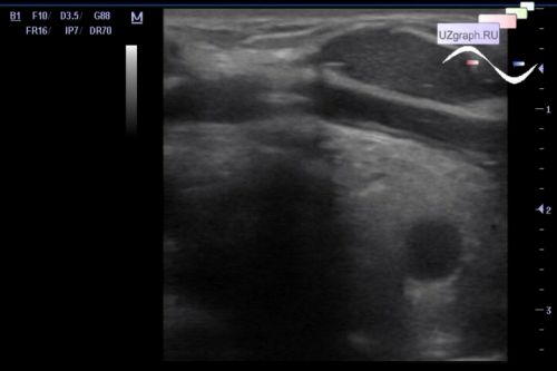
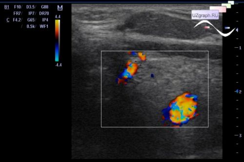
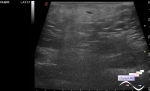
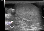
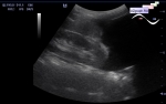
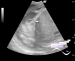
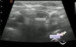
    A 14-year-old teenager undergoing medical screening at public clinic.

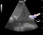
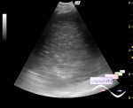
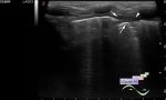
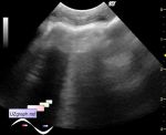
    On ultrasound of the thyroid gland, plus-tissue was visualized along its lower edge, it resembles a thymus gland in localization, but at this age thymus is usually no longer visualized.

    Dif. diagnosis: persistent thymus, lymphoma, etc.

    CT recommended.

    For those who do not believe that lymphoma may look like this, and are used to making a benign histological diagnosis by ultrasound in 100% of cases, the case of a colleague from the Netherlands - external link

    In addition, a small quote from a respected guide:

    "The thymus is a prominent normal anterior mediastinal structure in children up to 8 years of age. From age 2 to 8 years, the thymus remains a prominent sonographic landmark when scanning the mediastinum, even though it is less obvious on chest radiographs. Progressive fatty replacement makes the thymus blend with mediastinal fat and become sonographically invisible in older children and adults. Sonograpraphic visualization of the thymus in an adult suggests neoplastic disease."

    C.Rumack. Diagnostic ultrasound. Elsevier 2005 - p.619-621


 Random 
Ultrasound cases - Iliac kidney dystopia of abnormal shape
    

Ultrasound cases - Chest wound with barbed wire
    

Ultrasound cases - Thickening of the stomach wall
    

Ultrasound cases - Coccygeal cyst
    

Ultrasound cases - Twinkling artifacts plus
    

 Dictionary 
Ved

LVOT

CFM

RAP

ESD

Freeze

IVC

AC

FL

FVI


 Calculators 
Resistive index

TI-RADS

Teicholz systolic cardiac function

Myocardial mass

Body surface area


 Similar topics 
Ultrasound cases - Chylopericardium, lung atelectasis, cardiomegaly, ascites
    

Ultrasound cases - Diaphragmatic hernia, condition after surgery
    

Ultrasound cases - Hydrothorax
    

Ultrasound cases - One and a half kilograms of happiness
    

Ultrasound cases - Mirror artifact vs collapsed lung
    

Ultrasound cases - Air bronchogram
    

Ultrasound cases - Postoperative hydrothorax with sludge
    

Ultrasound cases - Pneumonia and hydrothorax on the left
    

Ultrasound cases - Diaphragmatic hernia, ectopic spleen & air...
    

Ultrasound cases - Diffusive-focal changes of the thymus