Log in    RU  


Main page :: Forum :: Ultrasound cases - Sonograms, cine-loops etc. ::

Goiter Hashimoto


Tags: Thyroid gland sonography, GE Vivid 3, Images, Video, Clinical report, Adult

   Posts
 
  Goiter Hashimoto
#1

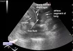
    The patient 43 years old came to control thyroid nodules.

    In the previous conclusion of the ultrasound several thyroid gland nodes are described, which are eventually attributed to thyroiditis.

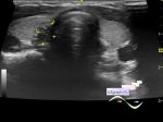
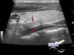
    On the current ultrasound, the thyroid gland has a clearly diffusely inhomogeneous structure; I did not see any sense in describing of some areas of heterogeneity which were visually separate from the general background. At the CFM blood flow is not enhanced. Presumably Hashimoto' s thyroiditis (autoimmune thyroiditis - AIT).

    external link

:: attachments(2) ::

:: file 1 ::

:: file 2 ::


 Random 
Ultrasound cases - Complete Duplicated Kidney, megaureters
    

Ultrasound cases - Cystic dysplasia, hematuria
    

Ultrasound cases - Condition after surgery - groin
    

Ultrasound cases - Hip dysplasia, subluxation
    

Ultrasound cases - One and a half kilograms of happiness
    

 Dictionary 
FL

MSD

PASP

PRF

PW

Ved

RAP

CRL

LVOT

B-mode


 Calculators 
Type of an infant hip joint

Thyroid volume

Volume (ellipse formula)

Estimated fetal weight by Ultrasound

Pressure gradient


 Similar topics 
Ultrasound cases - Tirads 5 multinodular
    

Ultrasound cases - Pediatric Tirads 5
    

Ultrasound cases - Tirads 5
    

Ultrasound cases - Thyroiditis 2
    

Ultrasound cases - Diffuse-focal thyroid changes, parathyroid glands adenomas
    

Ultrasound cases - Accessory Thyroid Lobe
    

Ultrasound cases - Area with hyperechoic dot inclusions
    

Ultrasound cases - Thyroiditis
    

Ultrasound cases - Hypoplasia of the left thyroid lobe
    

Ultrasound cases - Cystic-solid volume of the thyroid gland