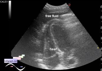
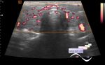
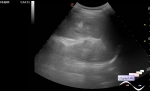
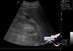
Condition after diaphragmatic hernia
Tags: Chest sonography, Abdomen sonography, Gastrointestinal sonography, Images, Video, Clinical report, Esaote MyLab 70, Pediatric
| Posts | |||
| Condition after diaphragmatic herni... | #1 |
| |||||
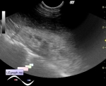
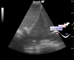
:: file 1 ::
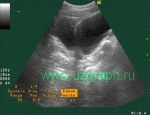
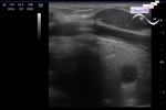
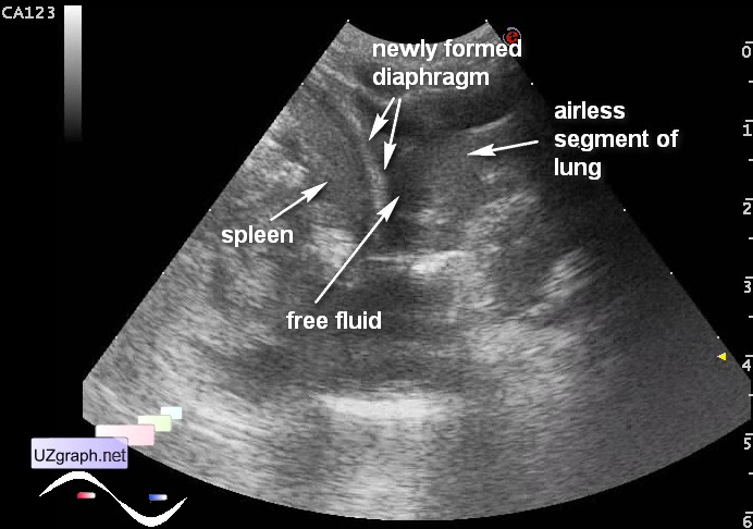 :: file 2 ::
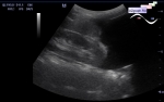
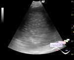
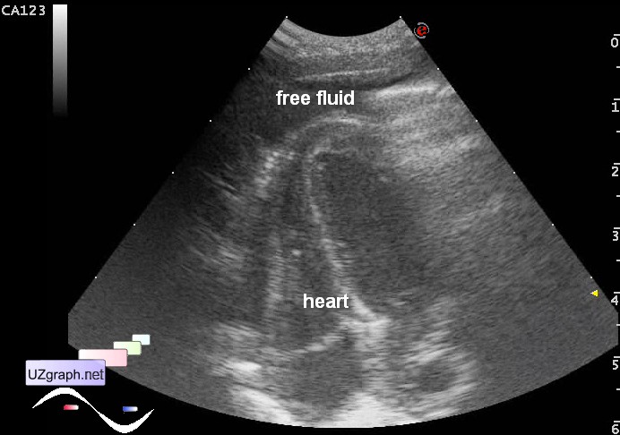 :: file 3 ::
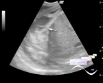
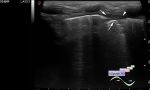
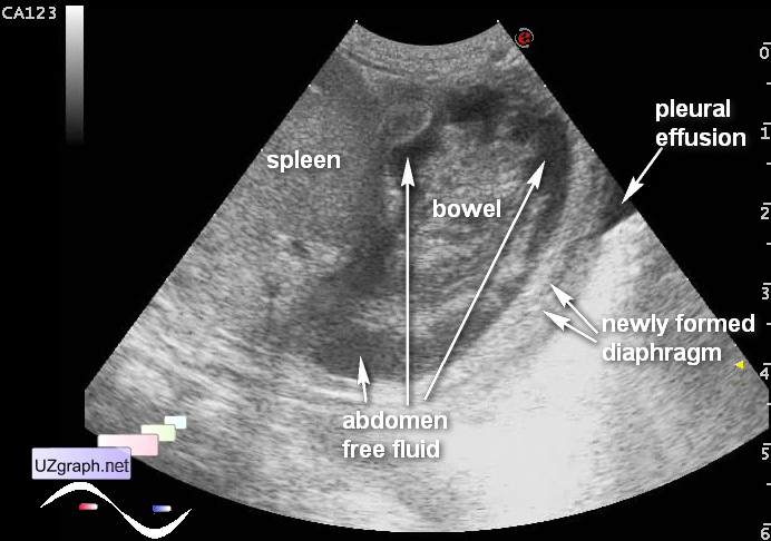 :: file 4 :: | |||||