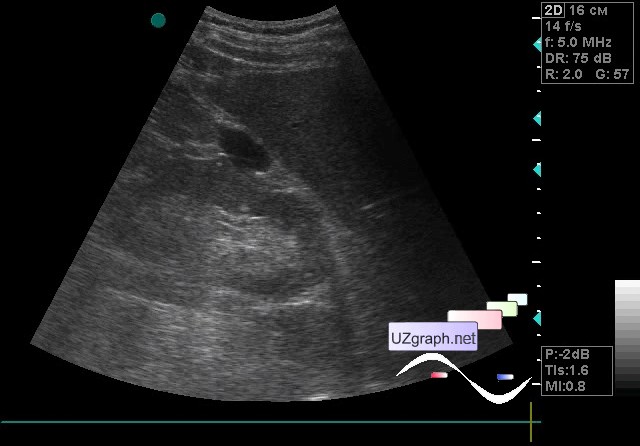
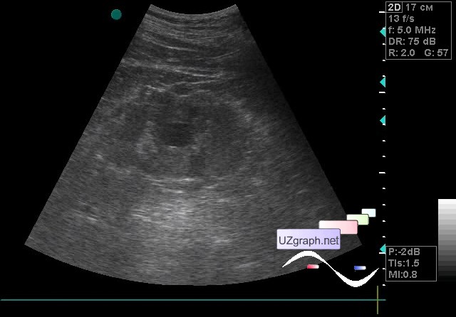
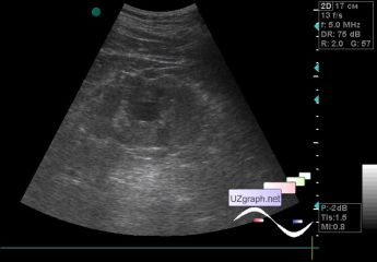
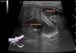
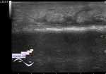
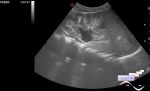
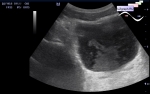
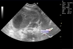
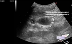
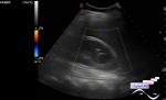
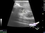
Log in    RU  


Main page :: Forum :: Ultrasound cases - Sonograms, cine-loops etc. ::

Kidney cysts


Tags: Urinary tract sonography, GE Vivid 3, Images, Video, Clinical report, Adult

   Posts
 
  Kidney cysts
#1

    A 68-year-old patient came for a kidney ultrasound.

    An anechoic lesion up to 2 cm is visualized on the anterior surface of the right kidney, on the CFM without blood flow (subcapsular cyst?).

    In the projection of the sinus of the left kidney, an anechoic lesion up to 2 cm is visualized, on the CFM without blood flow (sinus cyst?).

    external link

:: attachments(3) ::

:: file 1 ::

:: file 2 ::

:: file 3 ::


 Random 
Ultrasound cases - Patent ductus arteriosus(PDA)
    

Ultrasound cases - Purulent / Suppurative lymphadenitis
    

Ultrasound cases - Supposed renal hemangioma (angiomyolipoma)
    

Ultrasound cases - Lesions in the projection of the adrenal glands
    

Ultrasound cases - Pneumatosis intestinalis/ Necrotizing enterocolitis of newborns
    

 Dictionary 
CRL

LVESD

FL

SV

Doppler angle

EF

LVEDV

LN

M-mode

PSV


 Calculators 
Teicholz systolic cardiac function

Pulmonary artery pressure

Estimated fetal weight by Ultrasound

Type of an infant hip joint

Resistive index


 Similar topics 
Ultrasound cases - Autosomal recessive polycystic kidney disease (ARPKD)
    

Ultrasound cases - Bladder sludge
    

Ultrasound cases - Bright kidneys
    

Ultrasound cases - Ileostoma stenosis and nephropathy
    

Ultrasound cases - Renal pelvis clot
    

Ultrasound cases - Renal pelvis valve-stone
    

Ultrasound cases - Condition after treatment of a kidney cyst
    

Ultrasound cases - Hydronephrotic transformation & polypous bladder wall
    

Ultrasound cases - Pyelitis, cystitis
    

Ultrasound cases - Lesion in the projection of the adrenal gland 2