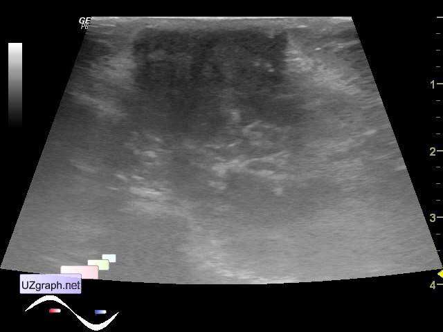
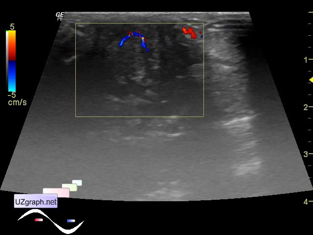
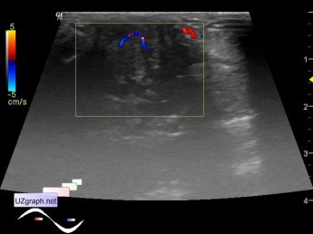
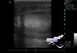
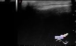
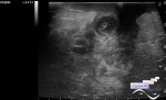
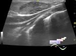
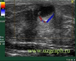
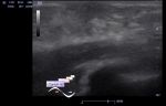
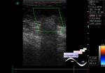
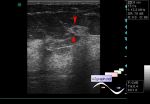
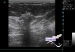
Log in    RU  


Main page :: Forum :: Ultrasound cases - Sonograms, cine-loops etc. ::

Gynecomastia


Tags: Breast sonography, Images, Video, Clinical report, GE Logiq P6, Pediatric

   Posts
 
  Gynecomastia
#1

    Boy 7 years old with no complaints, after the consultation of endocrinologist aimed to breast ultrasound.

    Visually nipples slightly raised on both sides.

    On US of subareolar area on both sides there was a symmetrical hypoechoic areas with increased blood flow on CFM.

:: attachments(3) ::

:: file 1 ::

:: file 2 ::

:: file 3 ::


 Random 
Ultrasound cases - Hydronephrosis & Ureterocele
    

Ultrasound cases - Chest wound with barbed wire
    

Ultrasound cases - Extra flow in the RV
    

Ultrasound cases - Free fluid and meniscuses
    

Ultrasound cases - Another polyp of the gallbladder
    

 Dictionary 
LVOT

EF

MSD

TAMEAN

PG

FNA

LVEDV

BPD

ESD

TVUS


 Calculators 
Body surface area

TI-RADS

Myocardial mass

Thyroid volume

Type of an infant hip joint


 Similar topics 
Ultrasound cases - Mastitis and cysts
    

Ultrasound cases - Neonatal mastitis
    

Ultrasound cases - Adolescent Gynecomastia
    

Ultrasound cases - Subacute mastitis (cyst with inflammation) in a teenager
    

Ultrasound cases - Breast Cysts
    

Ultrasound cases - Infant isolated thelarche
    

Ultrasound cases - Another Birads 4-5
    

Ultrasound cases - Star-like lesion
    

Ultrasound cases - Birads 4-5
    

Ultrasound cases - Breast lesions 3