Log in    RU  


Main page :: Forum :: Ultrasound cases - Sonograms, cine-loops etc. ::

Giant cyst of the right lateral canal


Tags: Female pelvis sonography, GE Logiq 400 MD, Clinical report, Pediatric

   Posts
 
  Giant cyst of the right lateral can...
#1

    A 12-year-old female patient came to the public clinic for a follow-up ultrasound; at birth, a right-sided nephrectomy was performed for a congenital tumor of the right kidney (cr). The last time she was seen by an oncologist a year ago, according to the medical data, no mts were found in the abdominal cavity.

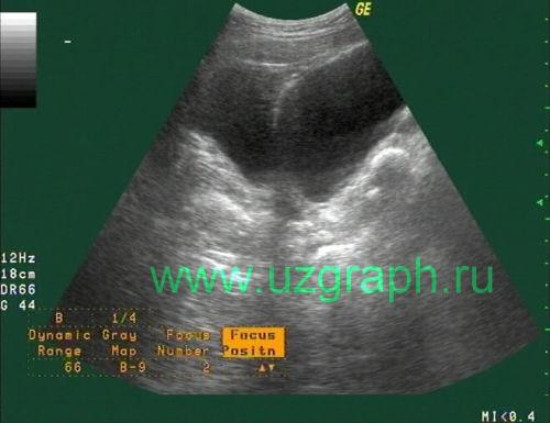
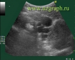
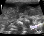
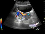
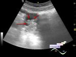
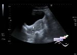
    On ultrasound, above the bladder a cyst is visualized, extending into the right lateral canal, with size approximately 16x6x10cm.

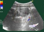
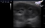
    During second ultrasound in other health care facilities, the cyst was preliminary diagnosed as an ovarian cyst, and on the next ultrasound the cyst was no longer visualized.


 Random 
Ultrasound cases - Onion rings sign
    

Ultrasound cases - Schlatter's disease
    

Ultrasound cases - Polyp of the gallbladder
    

Ultrasound cases - Cholelithiasis and hyperechoic gallbladder wall
    

Technical - Robosonography

 Dictionary 
SV

RAP

LVEDV

LMP

BSA

LVEDD

LN

Doppler angle

IVSd

Aliasing doppler artifact


 Calculators 
Type of an infant hip joint

Thyroid volume

Pressure gradient

Pulmonary artery pressure

Myocardial mass


 Similar topics 
Ultrasound cases - Nucks cyst
    

Ultrasound cases - Hydrosalpinx (saktosalpinx)
    

Ultrasound cases - Portal vein air in the newborn
    

Ultrasound cases - Ovarian teratoma
    

Ultrasound cases - Inguinal hernia with an ovary
    

Ultrasound cases - Right ovary changes
    

Ultrasound cases - Ovarian apoplexy 3
    

Ultrasound cases - Hemorrhagic ovarian cyst
    

Ultrasound cases - Patchy uterus
    

Ultrasound cases - Fibroid and nabothian cyst