Log in    RU  


Main page :: Forum :: Ultrasound cases - Sonograms, cine-loops etc. ::

Ovarian apoplexy 3


Tags: Female pelvis sonography, GE Logiq E, Clinical report, Pediatric

   Posts
 
  Ovarian apoplexy 3
#1

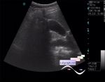
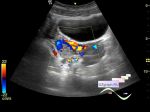
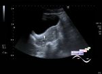
    A teenager in the public clinic was sent for a complex abdomen ultrasound with a diagnosis - examination - and complaints of recurrent abdominal pain.

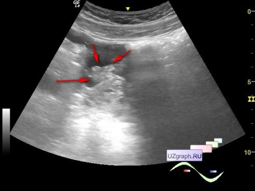
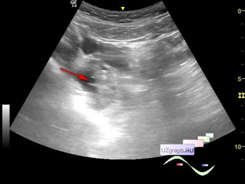
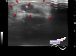
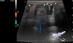
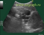
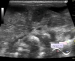
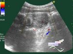
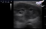
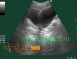
    On ultrasound in the right hypogastrium/small pelvis, the right ovary is visualized with an unclear contour (diff. diagnosis: apoplexy(ovarian rupture), etc.), surrounded by a small amount of free fluid (about 5 ml) and an echogenic lesion (diff. diagnosis: clot, omentum, etc.) .

    An emergency consultation with a gynecologist is recommended.


 Random 
Ultrasound cases - Double inferior vena cava
    

Ultrasound cases - ADPKD
    

Ultrasound cases - Strangulated inguinal hernia with necrosis/hematoma
    

Ultrasound cases - Intestinal intussusception, intestinal obstruction, ascites ...
    

Ultrasound cases - Incomplete membrane vs abnormal chord of LA
    

 Dictionary 
LN

PSV

GS

PRF

LMP

PASP

EF

FL

Color M-mode

CRL


 Calculators 
Gestational age by Ultrasound

Myocardial mass

Volume (ellipse formula)

TI-RADS

Resistive index


 Similar topics 
Ultrasound cases - Nucks cyst
    

Ultrasound cases - Hydrosalpinx (saktosalpinx)
    

Ultrasound cases - Portal vein air in the newborn
    

Ultrasound cases - Ovarian teratoma
    

Ultrasound cases - Inguinal hernia with an ovary
    

Ultrasound cases - Giant cyst of the right lateral canal
    

Ultrasound cases - Right ovary changes
    

Ultrasound cases - Hemorrhagic ovarian cyst
    

Ultrasound cases - Patchy uterus
    

Ultrasound cases - Fibroid and nabothian cyst