Log in    RU  


Main page :: Forum :: Ultrasound cases - Sonograms, cine-loops etc. ::

Patchy uterus


Tags: Female pelvis sonography, Esaote MyLab 70, Images, Video, Clinical report, Pediatric

   Posts
 
  Patchy uterus
#1

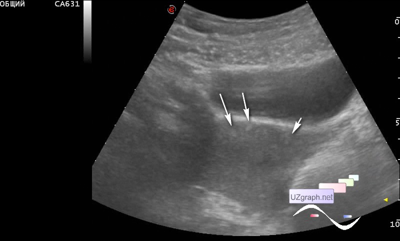
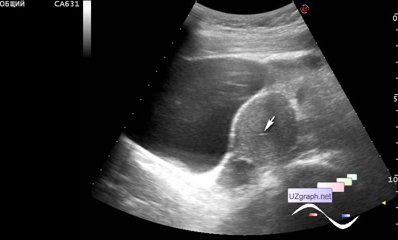
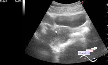
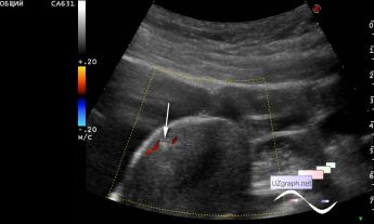
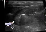
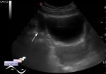
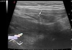
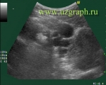
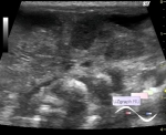
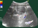
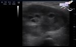
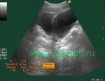
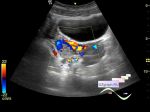
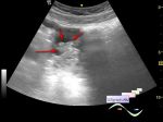
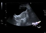
    A teenager with complaints of dysalgomenorrhea, day 6 of the cycle, by her words - menstruation is over.

    The size of the uterus, as in parous women. Myometrium is diffuse with hyperechoic lesions (endometriosis / adenomyosis?). In the upper and middle thirds of the uterine cavity - fluid, in the lower third - an echogenic lesion (clot?).

    At follow-up ultrasound, after 5 days, the “clot” and the fluid in the cavity disappeared, the uterus decreased to its normal size, but, as it says, “The longer they look, the more they'll find”. Upon careful viewing of the cine loop in the “off-line” mode - without a patient, new questions appeared - at the border of the lower and middle third, in the lateral parts of the uterus a linear structure (synechia?).

    external link

:: attachments(5) ::

:: file 1 ::

:: file 2 ::

:: file 3 ::

:: file 4 ::

:: file 5 ::


 Random 
Ultrasound cases - Focal thyroiditis plus
    

Ultrasound cases - Lesion in the projection of the anterior liver
    

Ultrasound cases - Yellow new white
    

Ultrasound cases - Hygromes of wrist
    

Ultrasound cases - If impossible but very much wanted then possible
    

 Dictionary 
HR

ESV

PG

LVOT

TV

LVESV

EDV

RAP

BSA

B-mode


 Calculators 
Resistive index

TI-RADS

Estimated fetal weight by Ultrasound

Thyroid volume

Myocardial mass


 Similar topics 
Ultrasound cases - Nucks cyst
    

Ultrasound cases - Hydrosalpinx (saktosalpinx)
    

Ultrasound cases - Portal vein air in the newborn
    

Ultrasound cases - Ovarian teratoma
    

Ultrasound cases - Inguinal hernia with an ovary
    

Ultrasound cases - Giant cyst of the right lateral canal
    

Ultrasound cases - Right ovary changes
    

Ultrasound cases - Ovarian apoplexy 3
    

Ultrasound cases - Hemorrhagic ovarian cyst
    

Ultrasound cases - Fibroid and nabothian cyst