Log in    RU  


Main page :: Forum :: Ultrasound cases - Sonograms, cine-loops etc. ::

Clinically suspected appendicitis 3


Tags: Female pelvis sonography, Images, Video, Clinical report, Esaote MyLab 70, Pediatric

   Posts
 
  Clinically suspected appendicitis 3
#1

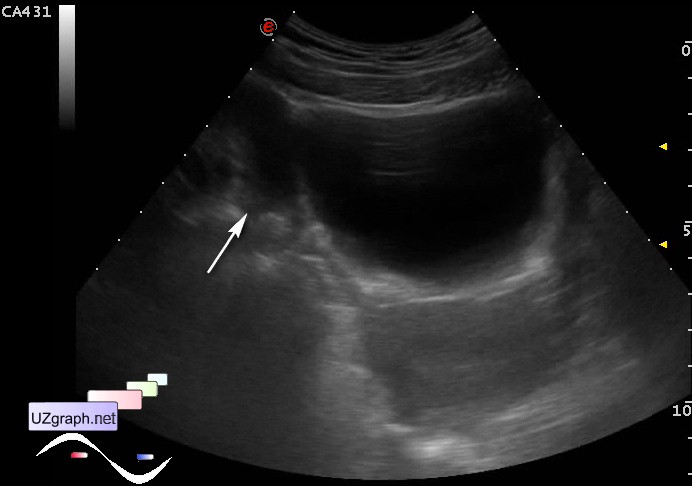
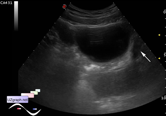
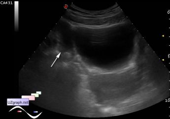
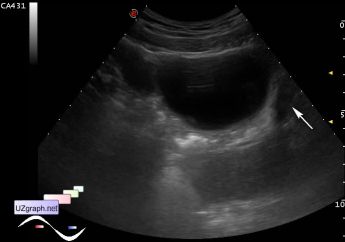
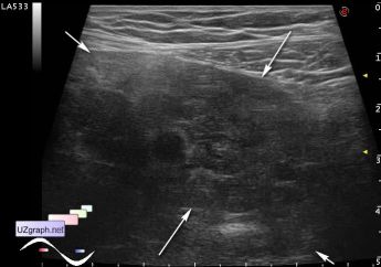
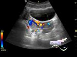
    Teenager girl with clinically suspected appendicitis, next day after admission, the previous ultrasound revealed appendix more than 6 mm in diameter. Assigned a control ultrasound.

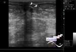
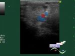
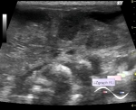
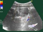
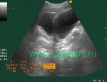
    At US appendix is not found, in the right iliac area there is a mass of solid-cystic structure up to 5.5 cm in length, on CFM with blood flow, presumably torsion of the right ovary.

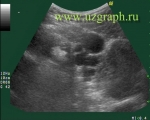
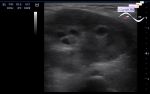
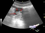
    Left ovary near to the uterus, normal sized and structure.

:: attachments(5) ::

:: file 1 ::

:: file 2 ::

:: file 3 ::

:: file 4 ::

:: file 5 ::


 Random 
Ultrasound cases - Incomplete testicular torsion
    

Ultrasound cases - Skin lesion in the projection of the surgical suture
    

Ultrasound cases - Testicular implant
    

Ultrasound cases - Subacute mastitis (cyst with inflammation) in a teenager
    

Ultrasound cases - Cyst-like lesion in the back of the neck in infant
    

 Dictionary 
ESV

HC

CO

ESD

TAMEAN

TV

MC

Qp-Qs

TAV

LVEDD


 Calculators 
Type of an infant hip joint

Volume (ellipse formula)

Estimated fetal weight by Ultrasound

Resistive index

Teicholz systolic cardiac function


 Similar topics 
Ultrasound cases - Nucks cyst
    

Ultrasound cases - Hydrosalpinx (saktosalpinx)
    

Ultrasound cases - Portal vein air in the newborn
    

Ultrasound cases - Ovarian teratoma
    

Ultrasound cases - Inguinal hernia with an ovary
    

Ultrasound cases - Giant cyst of the right lateral canal
    

Ultrasound cases - Right ovary changes
    

Ultrasound cases - Ovarian apoplexy 3
    

Ultrasound cases - Hemorrhagic ovarian cyst
    

Ultrasound cases - Patchy uterus