Log in    RU  


Main page :: Forum :: Ultrasound cases - Sonograms, cine-loops etc. ::

Nephrostomy problems


Tags: Urinary tract sonography, Images, Video, Clinical report, Esaote MyLab 70, Pediatric

   Posts
 
  Nephrostomy problems
#1

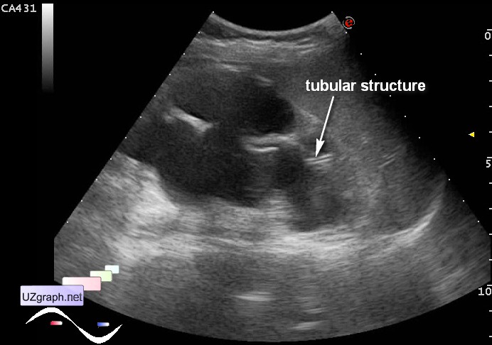
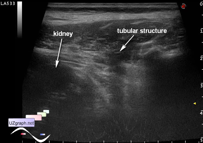
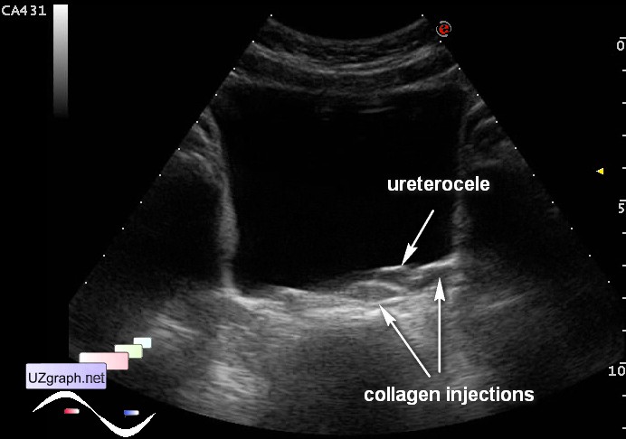
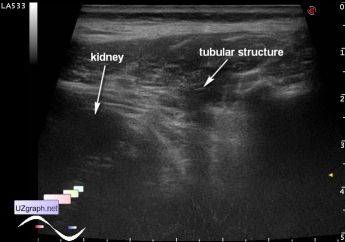
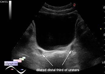
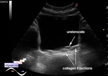
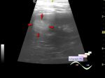
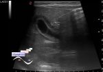
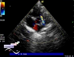
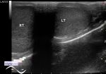
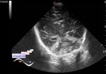
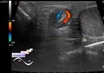
    Child 10 years old from the urology department with suspected nephrostomas malfunction (no urine through the nephrostomy during the day) is aimed to ultrasound.

    At ultrasound on the side of nephrostomy there is a severe hydronephrosis, the parenchyma is thinned to 2 mm. In the lumen of the upper renal calix the hyperechoic double linear structure is visualized type of a catheter - nephrostoma, with linear probe this structure is not visible, but such structure is visible in surrounding the upper third of the kidney tissues.

    In order to check the patency of nephrostomy with syringe was introduced saline in stoma and obtained the contrast effect (blizzard sign) in the renal pelvis.

    After the bladder visualized dilatation of the distal third of ureters, in the vesical triangle(Lieutaud triangle) on the side of hydronephrosis there is a small ureterocele and two small masses - due with several collagen injections in the past.

:: attachments(5) ::

:: file 1 ::

:: file 2 ::

:: file 3 ::

:: file 4 ::

:: file 5 ::


 Random 
Ultrasound cases - Redness of scrotum
    

Ultrasound cases - Perinephric lesion
    

Ultrasound cases - Goiter Hashimoto
    

Ultrasound cases - Hypertrophic pyloric stenosis
    

Ultrasound cases - Twinkling artifact
    

 Dictionary 
MSD

LVEDD

CRL

MPAP

PA

B-mode

VTI

HR

RVOT

CFM


 Calculators 
Qp:Qs

Resistive index

Pulmonary artery pressure

Teicholz systolic cardiac function

Volume (ellipse formula)


 Similar topics 
Ultrasound cases - Autosomal recessive polycystic kidney disease (ARPKD)
    

Ultrasound cases - Bladder sludge
    

Ultrasound cases - Bright kidneys
    

Ultrasound cases - Ileostoma stenosis and nephropathy
    

Ultrasound cases - Renal pelvis clot
    

Ultrasound cases - Renal pelvis valve-stone
    

Ultrasound cases - Condition after treatment of a kidney cyst
    

Ultrasound cases - Hydronephrotic transformation & polypous bladder wall
    

Ultrasound cases - Pyelitis, cystitis
    

Ultrasound cases - Lesion in the projection of the adrenal gland 2