Log in    RU  


Main page :: Forum :: Ultrasound cases - Sonograms, cine-loops etc. ::

PDA forth and back


Tags: Cardiac sonography(Echocardiography), Medison Sonoace R7, Images, Video, Clinical report, Pediatric

   Posts
 
  PDA forth and back
#1

    The child 1 year, has come on EchoCG on a screening.

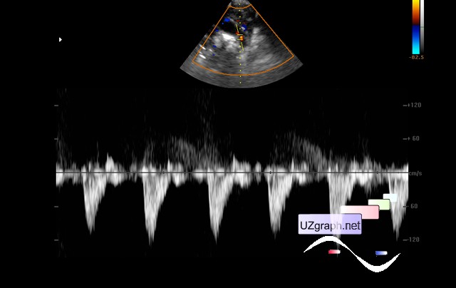
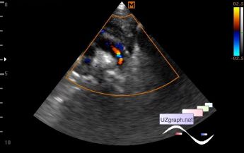
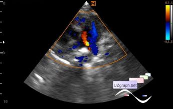
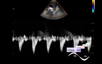
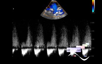
    In the projection of the descending part of the Ao arc , a retrograde flow with a Vmax up to 1.2 m / s to 3 mm in diameter. In the projection of the PA bifurcation is visualized retrograde flow up to 3 mm in diameter with a Vmax of up to 3 m / s (PDA with variable direction - bidirectional - of flow? Other?).

    Also, the PFO is visualized up to 5 mm (not in the images).

    external link

:: attachments(5) ::

:: file 1 ::

:: file 2 ::

:: file 3 ::

:: file 4 ::

:: file 5 ::


 Random 
Ultrasound cases - Record size appendicitis
    

Ultrasound cases - Strange RVOT
    

Ultrasound cases - Thyroid lesions
    

Ultrasound cases - Testicular hypoplasia, torsion of cord
    

On-call room - Interesting publication about neurosonography

 Dictionary 
ESD

IVC

TAMEAN

MC

TAV

Doppler angle

VTI

LVESV

EFW

LMP


 Calculators 
Pressure gradient

TI-RADS

Qp:Qs

Myocardial mass

Teicholz systolic cardiac function


 Similar topics 
Ultrasound cases - Bicuspid aortic valve 2
    

Ultrasound cases - Mitral valve cyst
    

Ultrasound cases - Electrode in the right heart after installing of heart pacemaker
    

Ultrasound cases - Chylopericardium, lung atelectasis, cardiomegaly, ascites
    

Ultrasound cases - Asymmetric LVH, aortic regurgitation
    

Ultrasound cases - Coronary fistula from LCA?
    

Ultrasound cases - Floating IAS aneurysm
    

Ultrasound cases - Aortic Stenosis
    

Ultrasound cases - Floating Accessory Pulmonary Valve Tissue
    

Ultrasound cases - Hyperechoic linear structure in the left atrium