Log in    RU  


Main page :: Forum :: Ultrasound cases - Sonograms, cine-loops etc. ::

Something on the neck


Tags: Soft tissues sonography, Images, Video, Clinical report, Esaote MyLab 70, Pediatric

   Posts
 
  Something on the neck
#1

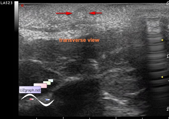
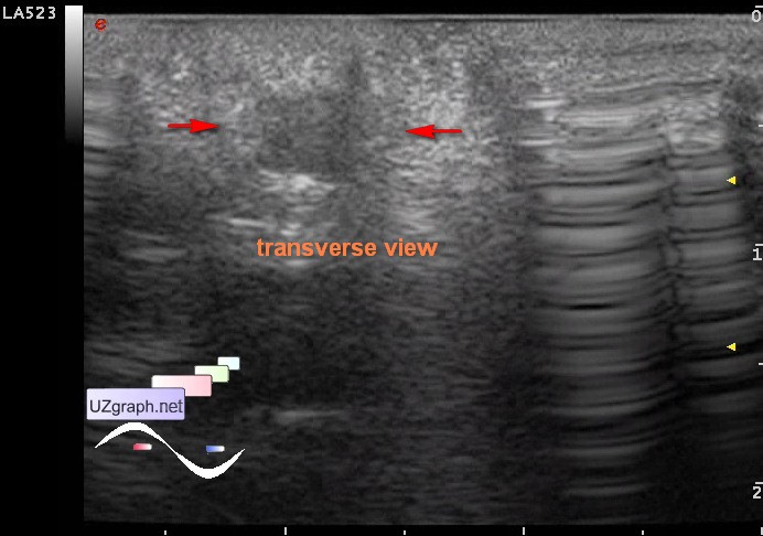
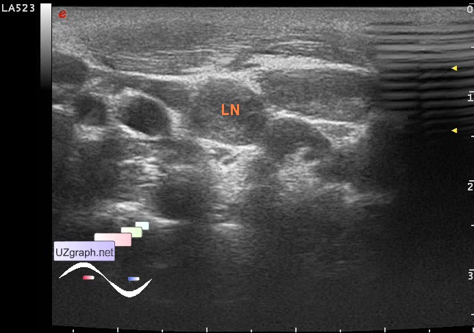
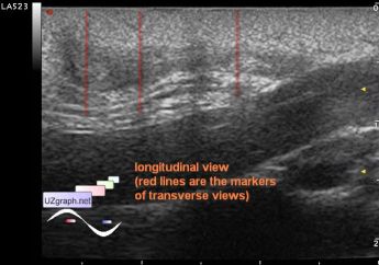
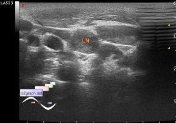
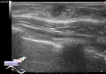
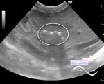
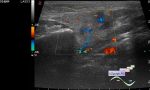
    A child 4 years-old with mental retardation, from birth observed with mass looks like polyp on the side of the neck, painless when touching by probe, bendable, aimed to ultrasound before surgery.

    At US (mass pressed to the neck by probe) the mass is heteroechoic, has 2-layers: hyperechoic outer layer and an/ hypoechoic inner layer, small hypoechoic lesions inside can't be excluded, at CFM/ DPD there is no true evidence of blood flow, size of the mass-body (part of mass which above the level of the body surface) is approximately 2,5x1cm, the hypoechoic leg of the mass up to 3 mm in diameter goes to the subcutaneous fat layer up to the level of muscle and then turns in the direction of the trachea, the further course of the leg can not be traced (dif.diagnosis: teratoma/dermoid, cartilaginous exostosis, etc.).

    On both sides of the neck there is a few enlarged LN with blood flow at CFM (dif.diagnosis: lymphadenitis, lymphadenopathy).

    external link

:: attachments(10) ::

:: file 1 ::

:: file 2 ::

:: file 3 ::

:: file 4 ::

:: file 5 ::

:: file 6 ::

:: file 7 ::

:: file 8 ::

:: file 9 ::

:: file 10 ::


 Random 
Ultrasound cases - Acute cervical lymphadenitis
    

Ultrasound cases - The effect of extreme
    

Ultrasound cases - Mirror artifact vs collapsed lung
    

Scientific - ASE Webinars about COVID-19

Ultrasound cases - Hip dysplasia, subluxation
    

 Dictionary 
TV

B-mode

CRL

SF

AC

Qp-Qs

TI-RADS

PASP

LN

TAV


 Calculators 
Resistive index

TI-RADS

Thyroid volume

Gestational age by Ultrasound

Teicholz systolic cardiac function


 Similar topics 
Ultrasound cases - Penetrating injury of the femur
    

Ultrasound cases - Cephalohematoma
    

Ultrasound cases - Urachus fistula
    

Ultrasound cases - Cranial mass
    

Ultrasound cases - Pilonidal coccyx cyst
    

Ultrasound cases - Palm fistula, in search of pencil lead
    

Ultrasound cases - Postoperative abdominal wall fistula
    

Ultrasound cases - Nucks cyst
    

Ultrasound cases - Paraproctitis
    

Ultrasound cases - Submandibular area lesion