One kidney two uteri
Tags: Urinary tract sonography, Female pelvis sonography, Images, Video, Clinical report, Esaote MyLab 70, Pediatric
| Posts | |||
| One kidney two uteri | #1 |
| |||||
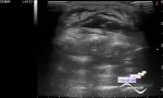
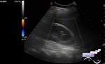
:: file 1 ::
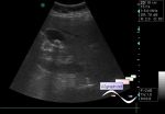
 :: file 2 ::
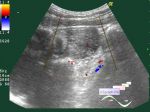
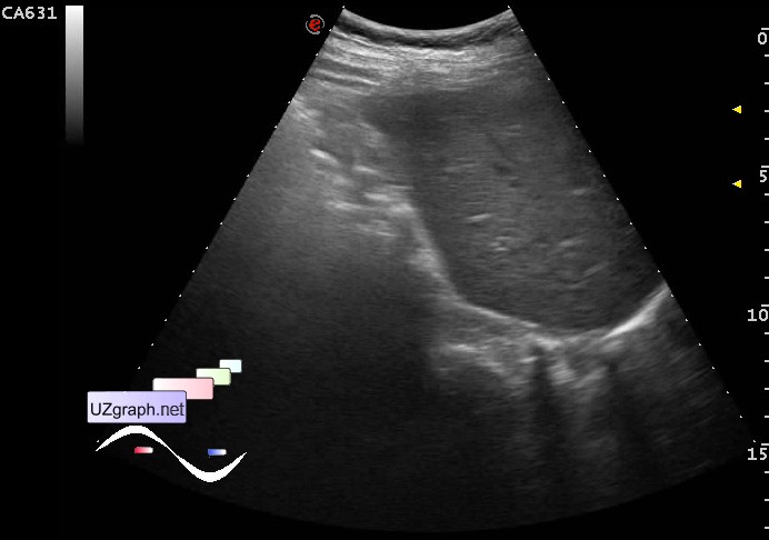
 :: file 3 ::
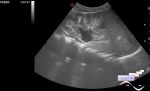
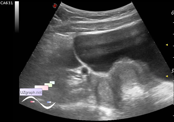
 :: file 4 ::
 :: file 5 ::
 :: file 6 ::
 :: file 7 ::
 :: file 8 :: | |||||