Log in    RU  


Main page :: Forum :: Ultrasound cases - Sonograms, cine-loops etc. ::

Ovarian teratoma


Tags: Female pelvis sonography, GE Logiq 400 MD, Clinical report, Pediatric

   Posts
 
  Ovarian teratoma
#1

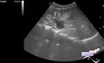
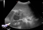
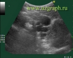
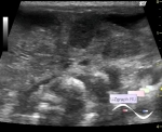
    A teenage girl came for a routine ultrasound scan at a public clinic, an accidental finding.

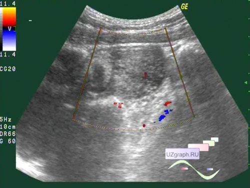
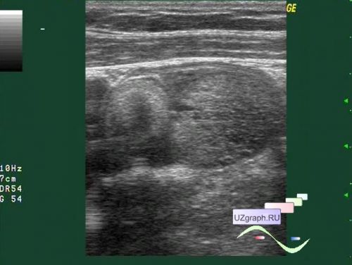
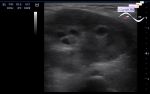
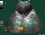
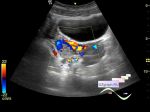
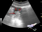
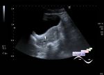
    In one of the ovaries, 2 hyperechoic lesions are visualized, the first resembles a horseshoe in shape, the second is of an irregular shape with an unclear contour; on the CFM, single locus of blood flow are mapped in the second lesion (most likely teratoma / teratomas).


 Random 
Ultrasound cases - White/ bright/ hyperechoic kidney symptom
    

Ultrasound cases - Hydrocele & cryptorchidism
    

Ultrasound cases - Subcutaneous shoulder lesion
    

Ultrasound cases - Condition after choledochal cyst removal
    

Ultrasound cases - Hydrothorax and abdominal syndrome
    

 Dictionary 
MC

LVOT

SF

IVSd

ESV

MSD

EF

TI-RADS

PVR

EDD


 Calculators 
Pressure gradient

Type of an infant hip joint

Qp:Qs

Thyroid volume

Resistive index


 Similar topics 
Ultrasound cases - Nucks cyst
    

Ultrasound cases - Hydrosalpinx (saktosalpinx)
    

Ultrasound cases - Portal vein air in the newborn
    

Ultrasound cases - Inguinal hernia with an ovary
    

Ultrasound cases - Giant cyst of the right lateral canal
    

Ultrasound cases - Right ovary changes
    

Ultrasound cases - Ovarian apoplexy 3
    

Ultrasound cases - Hemorrhagic ovarian cyst
    

Ultrasound cases - Patchy uterus
    

Ultrasound cases - Fibroid and nabothian cyst