Log in    RU  


Main page :: Forum :: Ultrasound cases - Sonograms, cine-loops etc. ::

Inguinal hernia with an ovary


Tags: Soft tissues sonography, Female pelvis sonography, Mindray M5, Clinical report, Pediatric

   Posts
 
  Inguinal hernia with an ovary
#1

    Parents of a 6-month-old girl turned to the surgeon in the public clinic with complaints about the bulging in the groin that appeared in the child the other day after the massage. The surgeon sent them for an urgent ultrasound.

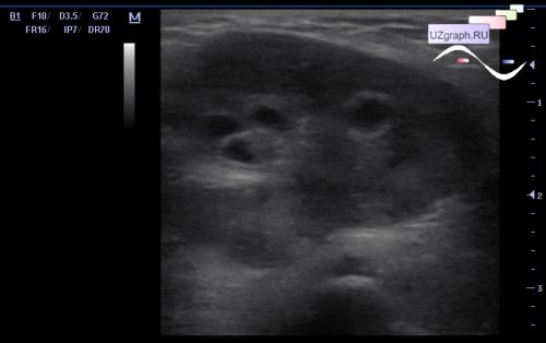
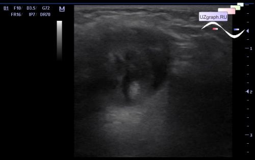
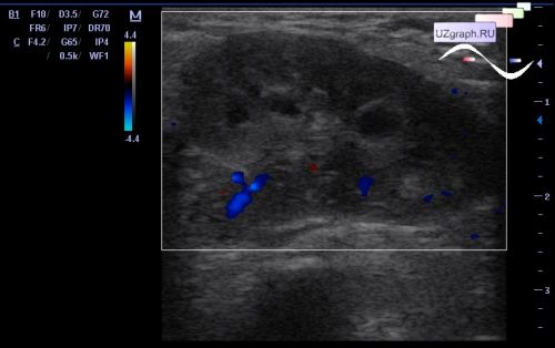
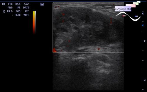
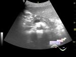
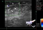
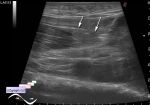
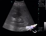
    On ultrasound in the groin, a lesion with an echo-pattern resembling a kidney is visualized, solid with the an/hypoechoic rounded inclusions with an echogenic rim, with no blood flow at CFM and PD, two vessels are visualized at the gate of the lesion at CFM, at PW the arterial spectrum with RI = 0.8. The bladder and both kidneys are without features.

    A strangulated inguinal hernia was suspected and the child was hospitalized. According to a colleague, the solid lesion in the groin turned out to be an ovary.


 Random 
Ultrasound cases - Haemorrhagic ovarian cyst
    

Ultrasound cases - If impossible but very much wanted then possible
    

Ultrasound cases - Thread-like structure in RA
    

Ultrasound cases - Clinic of appendicitis or ascites, peritoneum disseminates & hernia
    

Ultrasound cases - Progressive giant intussusception
    

 Dictionary 
Color M-mode

Freeze

PADP

LVOT

CW

Qp-Qs

HC

PSV

BSA

EFW


 Calculators 
Resistive index

Body surface area

Gestational age by Ultrasound

Pulmonary artery pressure

Pressure gradient


 Similar topics 
Ultrasound cases - Penetrating injury of the femur
    

Ultrasound cases - Cephalohematoma
    

Ultrasound cases - Urachus fistula
    

Ultrasound cases - Cranial mass
    

Ultrasound cases - Pilonidal coccyx cyst
    

Ultrasound cases - Palm fistula, in search of pencil lead
    

Ultrasound cases - Postoperative abdominal wall fistula
    

Ultrasound cases - Nucks cyst
    

Ultrasound cases - Paraproctitis
    

Ultrasound cases - Submandibular area lesion