Log in    RU  


Main page :: Forum :: Ultrasound cases - Sonograms, cine-loops etc. ::

Portal vein spontaneous enhancement (mononucleosis)


Tags: Abdomen sonography, GE Logiq E, Clinical report, Pediatric

   Posts
 
  Portal vein spontaneous enhancement...
#1

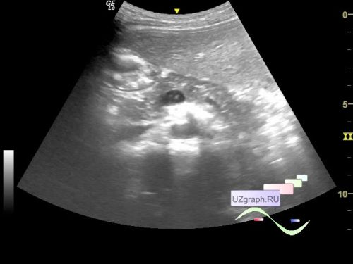
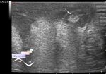
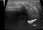
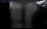
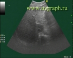
    A 7-year-old girl is undergoing treatment for infectious mononucleosis and was sent for a follow-up abdominal and kidneys ultrasound; a previous ultrasound revealed hepatomegaly.

    On the current ultrasound, the liver is normal in size but in the portal vein in B-mode a symptom of spontaneous enhancement is visualized (atypical mononuclear cells?).


 Random 
Ultrasound cases - Subluxation of the hip joint (type III Graf)
    

Ultrasound cases - Old hydrocele, testicular changes
    

Ultrasound cases - Pendulum-like intestine peristalsis
    

Ultrasound cases - Only the questions
    

Ultrasound cases - PDA 7yo
    

 Dictionary 
IVSd

Aliasing doppler artifact

IVC

GS

LVOT

PSV

PADP

TAMEAN

FVI

M-mode


 Calculators 
Teicholz systolic cardiac function

Volume (ellipse formula)

Estimated fetal weight by Ultrasound

Type of an infant hip joint

Pressure gradient


 Similar topics 
Ultrasound cases - Hirschsprung disease on ultrasound
    

Ultrasound cases - Intestinal Infiltrate
    

Ultrasound cases - Abdominal cryptorchidism
    

Ultrasound cases - Diffuse heterogeneous liver
    

Ultrasound cases - Postoperative abdominal wall fistula
    

Ultrasound cases - Stump appendicitis
    

Ultrasound cases - Liver scar after resection
    

Ultrasound cases - Subdiaphragmatic movable fluid collection (UFO)
    

Ultrasound cases - Lesion of the spleen or the case when it is easy to not notice
    

Ultrasound cases - I’m normal but others ...