Log in    RU  


Main page :: Forum :: Ultrasound cases - Sonograms, cine-loops etc. ::

Prepatellar bursitis


Tags: Soft tissues sonography, Musculoskeletal sonography, Mindray M5, Images, Video, Clinical report, Pediatric

   Posts
 
  Prepatellar bursitis
#1

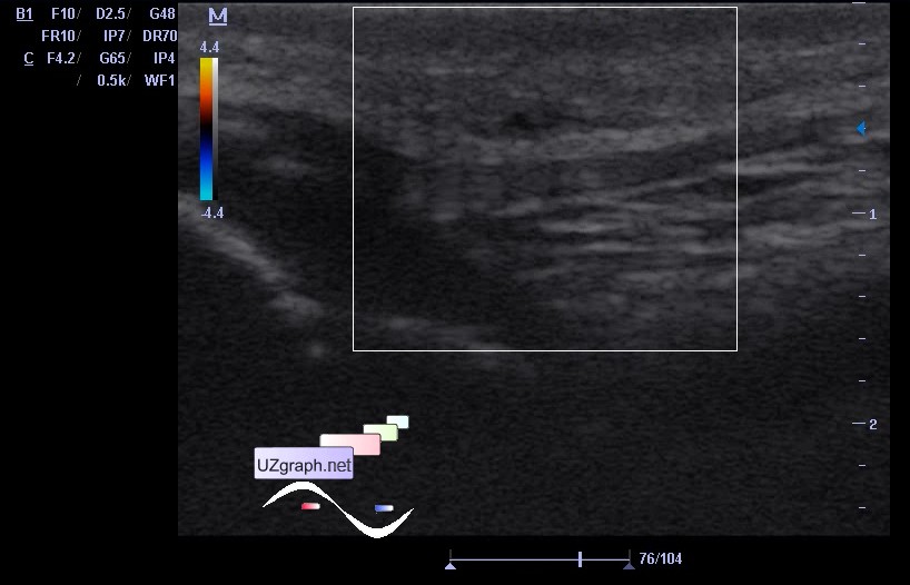
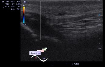
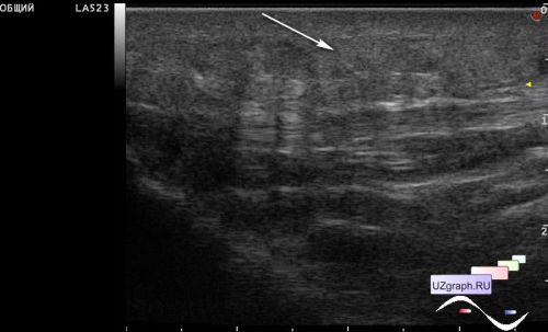
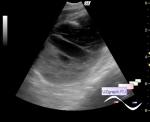
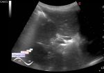
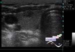
    A child of 5 years old with complains of periodic knee pain that appeared several days ago, which occurs when touching the front surface of the knee and while walking pain which does not limit mobility in the knee joint.

    Consulted by a surgeon and orthopedic surgeon, directed to an ultrasound scan with suspected inflammatory process of the corresponding knee.

    Upon visual inspection, both knees on the front surface, the patient is more likely to have thickened rough-skin as a skin corn.

    The child does not notice a pronounced pain when touching the zone of interest.

    At ultrasound scan visualised an asymmetry of skin thickness and subcutaneus fat: right(affected side) > left, marked heterogeneity of the skin/subcutaneus fat echo-structure on the right, increased blood flow by venous type at spectral doppler.

    PS. files 1-3 right knee, file 4 - left knee.

    external link

:: attachments(4) ::

:: file 1 ::

:: file 2 ::

:: file 3 ::

:: file 4 ::

  08:55 21-05-2022
  Prepatellar bursitis (teenager)
#2

    A 14-year-old female teenager who returned from a long hike, with complaints of a dense, painful while walking, mass in the front surface of the right knee came to the ED of the Children's City Clinical Hospital and was urgently sent for an ultrasound scan.

    On ultrasound, no mass was found, in the projection of complaints, a slightly thickened, pronounced heterogeneous, increased echogenicity subcutaneous fatty tissue along the anterior surface of the right knee (prepatellar bursa, prepatellaris subcutanea) is visualized.


 Random 
Ultrasound cases - Ectopic thymus
    

Ultrasound cases - Pulmonary stenosis
    

Ultrasound cases - Another polyp of the gallbladder
    

Ultrasound cases - Riedels lobe vs diaphragmatic hernia
    

Ultrasound cases - Thickening of the stomach wall
    

 Dictionary 
PRF

IVC

SF

BPD

PA

PG

HR

PSV

LMP

PASP


 Calculators 
Volume (ellipse formula)

Teicholz systolic cardiac function

Pulmonary artery pressure

Thyroid volume

Body surface area


 Similar topics 
Ultrasound cases - Penetrating injury of the femur
    

Ultrasound cases - Cephalohematoma
    

Ultrasound cases - Urachus fistula
    

Ultrasound cases - Cranial mass
    

Ultrasound cases - Pilonidal coccyx cyst
    

Ultrasound cases - Palm fistula, in search of pencil lead
    

Ultrasound cases - Postoperative abdominal wall fistula
    

Ultrasound cases - Nucks cyst
    

Ultrasound cases - Paraproctitis
    

Ultrasound cases - Submandibular area lesion