Log in    RU  


Main page :: Forum :: Ultrasound cases - Sonograms, cine-loops etc. ::

Thyroid lesions 3


Tags: Thyroid gland sonography, GE Vivid 3, Images, Video, Clinical report, Adult

   Posts
 
  Thyroid lesions 3
#1

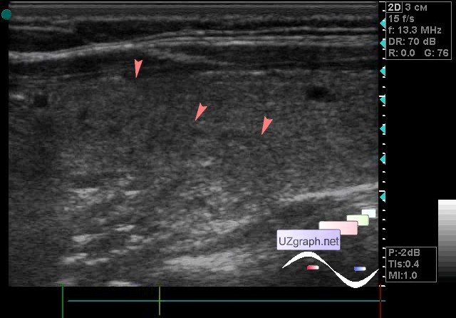
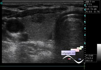
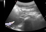
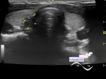
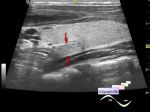
    A patient 36 years old came to the control of the thyroid ultrasound with a diagnosis of a multinodular goiter, in the previous conclusion of the ultrasound scan 4 nodes were described, 2 nodes in each lobe.

    On the current ultrasound in the right lobe a single predominantly hypoechoic lesion of an oval shape up to 7 mm(colloid node? adenoma? other?), at CFM without blood flow, as well as a region of reduced echogenicity up to 2.5 mm (follicle?) are visualized; in the left lobe a single follicle up to 2.5 mm and an area of heterogeneity in the posterior parts of the lobe approximately 2.5 x 0.5 x 1.5 cm (thyroiditis?) are visualized.

:: attachments(3) ::

:: file 1 ::

:: file 2 ::

:: file 3 ::


 Random 
Admin room - We're open

Ultrasound cases - Breast lesions 3
    

Ultrasound cases - The worse is better
    

Ultrasound cases - Stump appendicitis
    

Ultrasound cases - Post-traumatic ankle arthritis
    

 Dictionary 
IVC

PG

HC

RAP

FVI

ESV

AC

Ved

EDD

TV


 Calculators 
Type of an infant hip joint

TI-RADS

Teicholz systolic cardiac function

Gestational age by Ultrasound

Myocardial mass


 Similar topics 
Ultrasound cases - Tirads 5 multinodular
    

Ultrasound cases - Pediatric Tirads 5
    

Ultrasound cases - Tirads 5
    

Ultrasound cases - Thyroiditis 2
    

Ultrasound cases - Diffuse-focal thyroid changes, parathyroid glands adenomas
    

Ultrasound cases - Accessory Thyroid Lobe
    

Ultrasound cases - Area with hyperechoic dot inclusions
    

Ultrasound cases - Thyroiditis
    

Ultrasound cases - Hypoplasia of the left thyroid lobe
    

Ultrasound cases - Cystic-solid volume of the thyroid gland