Log in    RU  


Main page :: Forum :: Ultrasound cases - Sonograms, cine-loops etc. ::

Hemangiomatosis of mammary gl. and birads 4-5


Tags: Breast sonography, GE Vivid 3, Images, Video, Clinical report, Adult

   Posts
 
  Hemangiomatosis of mammary gl. and ...
#1

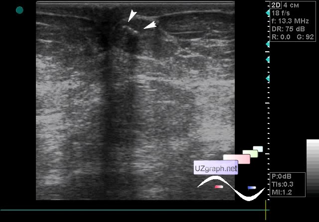
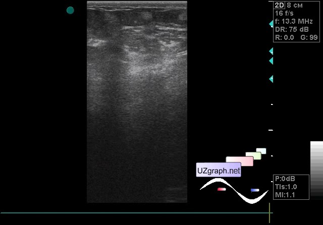
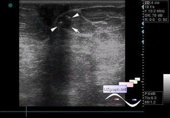
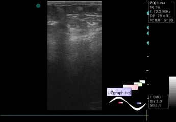
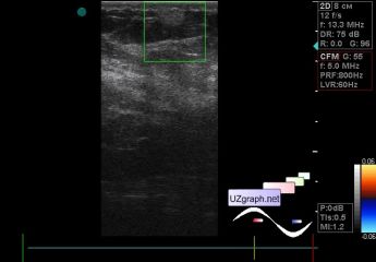
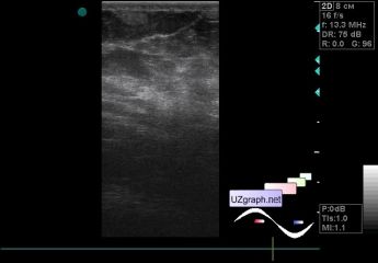
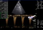
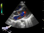
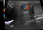
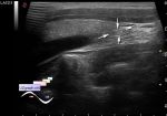
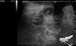
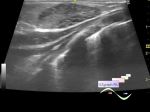
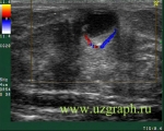
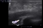
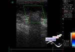
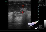
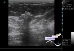
    A 76-year-old patient came to a screening ultrasound of the mammary glands, glands are large, according to her words there was no earlier breast ultrasound scans, but a diagnosis of lipomatosis was made, in particular, the patient had a large mass in the forearm and in the mammary glands she found several mass at palpation.

    On ultrasound, both mammary glands have a diffusely heterogeneous structure due to multiple hyperechoic inclusions of various shapes and sizes (dozens in each gland - hemangiomas?), Without blood flow at CFM. On their background, I hardly noticed in the nipple area of the one of the mammary glands a chain of hyperechoic microinclusions(microcalcinates?), I looked closely and they were along the contour of a hypoechoic rounded inclusion, on the CFM without blood flow (birads 4-5?).

    external link

:: attachments(6) ::

:: file 1 ::

:: file 2 ::

:: file 3 ::

:: file 4 ::

:: file 5 ::

:: file 6 ::


 Random 
Ultrasound cases - Subtotal encephalomalacia
    

Ultrasound cases - Cholelithiasis
    

Ultrasound cases - Yellow new white
    

Admin room - Feedback - an error was found

Ultrasound cases - Diaphragmatic hernia, condition after surgery
    

 Dictionary 
EFW

Aliasing doppler artifact

PG

PADP

EDD

B-mode

TVUS

TAMEAN

TV

MC


 Calculators 
Teicholz systolic cardiac function

Resistive index

Pulmonary artery pressure

Thyroid volume

TI-RADS


 Similar topics 
Ultrasound cases - Mastitis and cysts
    

Ultrasound cases - Neonatal mastitis
    

Ultrasound cases - Adolescent Gynecomastia
    

Ultrasound cases - Subacute mastitis (cyst with inflammation) in a teenager
    

Ultrasound cases - Breast Cysts
    

Ultrasound cases - Infant isolated thelarche
    

Ultrasound cases - Another Birads 4-5
    

Ultrasound cases - Star-like lesion
    

Ultrasound cases - Birads 4-5
    

Ultrasound cases - Breast lesions 3