Log in    RU  


Main page :: Forum :: Ultrasound cases - Sonograms, cine-loops etc. ::

Suspected gastric ulcer


Tags: Gastrointestinal sonography, Images, Video, Clinical report, Esaote MyLab 70, Pediatric

   Posts
 
  Suspected gastric ulcer
#1

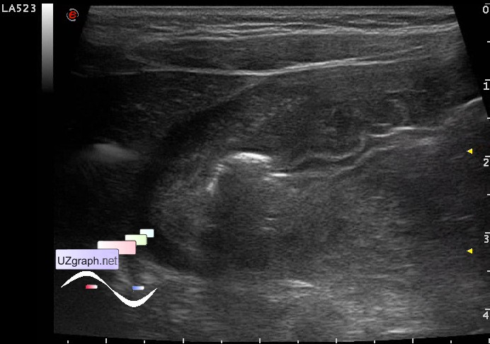
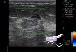
    A child of 4 years old with suspected appendicitis.

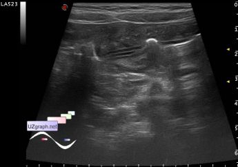
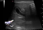
    Vermiform appendix unremarkable.

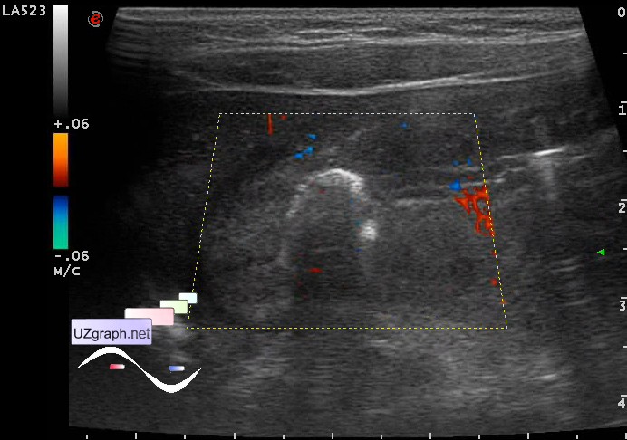
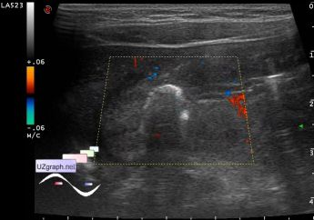
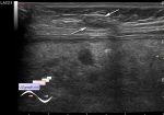
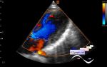
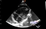
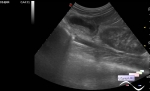
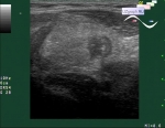
    The pyloric of stomach closed with a thickened wall up to 8 mm (gastritis?), at CFM with the increased blood flow, along the front wall there is a crater-like hyperechoic lesion up to 8 mm in diameter (ulcer?)

    external link

:: attachments(5) ::

:: file 1 ::

:: file 2 ::

:: file 3 ::

:: file 4 ::

:: file 5 ::

  11:14 13-04-2016
 
#2

    Diagnosis was confirmed with a correction - antral(prepyloric) gastric ulcer.


 Random 
Ultrasound cases - Area with hyperechoic dot inclusions
    

Ultrasound cases - Hyperechoic segment of the kidney
    

Ultrasound cases - Lesion of the front wall of chest
    

Ultrasound cases - Acute orchoepididymitis secondary to torsion ...
    

On-call room - MRI communism

 Dictionary 
CFM

TVUS

Qp-Qs

Color M-mode

PW

LVPWd

MC

ESD

DPD

Freeze


 Calculators 
Estimated fetal weight by Ultrasound

Teicholz systolic cardiac function

Qp:Qs

Body surface area

Gestational age by Ultrasound


 Similar topics 
Ultrasound cases - Ileotiphlitis and mesadenitis
    

Ultrasound cases - Acute Appendicitis, periappendicular infiltrate
    

Ultrasound cases - Megacolon
    

Ultrasound cases - Necrotizing Enterocolitis
    

Ultrasound cases - Periappendicular Abscess
    

Ultrasound cases - Acute Appendicitis
    

Ultrasound cases - 1 kilogram of happiness
    

Ultrasound cases - Appendicolith
    

Ultrasound cases - Hollow organ lesion sign, "Lost in Translation"
    

Ultrasound cases - Pneumatosis intestinalis/ Necrotizing enterocolitis of newborns