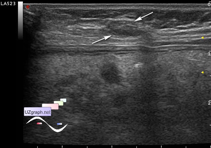
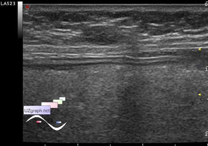
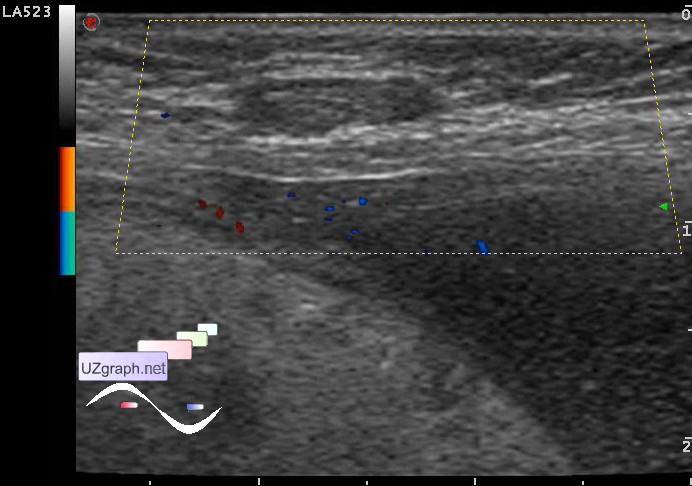
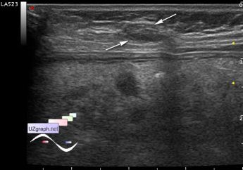
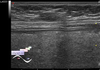
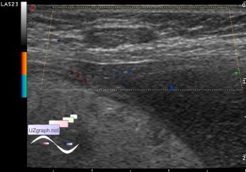
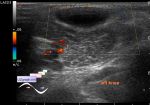
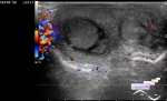
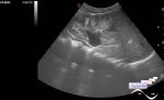
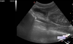
Log in    RU  


Main page :: Forum :: Ultrasound cases - Sonograms, cine-loops etc. ::

Linea alba hernia vs lipoma part trois


Tags: Soft tissues sonography, Images, Video, Clinical report, Esaote MyLab 70, Pediatric

   Posts
 
  Linea alba hernia vs lipoma part tr...
#1

    Child 2 years old after surgeons consultation aimed to ultrasound with diagnosis - hernia of the linea alba / lipoma.

    Palpable just above the umbilicus on the center line of the abdomen there is a solid lesion up to 1 cm.

    At US in this projection there is an oval shaped hypoechoic lesion with lateral shadows and no evidence of blood flow at CFM(DPD) - presumably lipoma.

    Previous episodes:

    https://m.en.uzgraph.ru/forum/...

    https://m.en.uzgraph.ru/forum/...

    external link

:: attachments(4) ::

:: file 1 ::

:: file 2 ::

:: file 3 ::

:: file 4 ::


 Random 
Ultrasound cases - Subdiaphragmatic movable fluid collection (UFO)
    

Ultrasound cases - Coronary fistula from LCA?
    

Ultrasound cases - Subdural hemorrhage 2
    

Ultrasound cases - Meconium peritonitis and ascites
    

Scientific - Playlist Chest ultrasound

 Dictionary 
LVEDV

Aliasing doppler artifact

ESD

AC

Color M-mode

ESV

PG

SV

VTI

M-mode


 Calculators 
Resistive index

Qp:Qs

TI-RADS

Thyroid volume

Pressure gradient


 Similar topics 
Ultrasound cases - Penetrating injury of the femur
    

Ultrasound cases - Cephalohematoma
    

Ultrasound cases - Urachus fistula
    

Ultrasound cases - Cranial mass
    

Ultrasound cases - Pilonidal coccyx cyst
    

Ultrasound cases - Palm fistula, in search of pencil lead
    

Ultrasound cases - Postoperative abdominal wall fistula
    

Ultrasound cases - Nucks cyst
    

Ultrasound cases - Paraproctitis
    

Ultrasound cases - Submandibular area lesion