Log in    RU  


Main page :: Forum :: Ultrasound cases - Sonograms, cine-loops etc. ::

Cysts and milk ducts


Tags: Breast sonography, GE Vivid 3, Images, Video, Clinical report, Adult

   Posts
 
  Cysts and milk ducts
#1

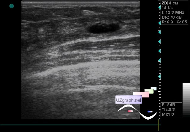
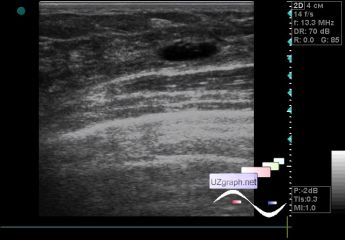
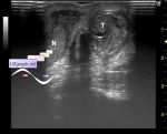
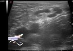
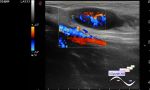
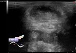
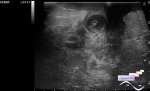
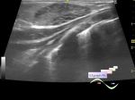
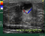
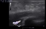
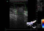
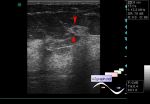
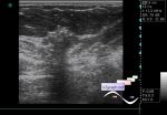
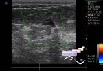
    A 44-year-old patient came for an ultrasound of the mammary glands; earlier, according to her, cysts were found in one gland.

    On ultrasound, in the external parts of the left gland, two cysts are visualized(fig.1-3), one of them with hyperechoic point inclusions, on the CFM without blood flow. In the retroareolar region the dilated milk ducts(mammary duct ectasia, fig.4) up to 4 mm, also with echogenic point inclusions, during compression of this region the patient noted discomfort (mastitis? mastopathy?). Milk ducts in another gland up to 3 mm.

:: attachments(5) ::

:: file 1 ::

:: file 2 ::

:: file 3 ::

:: file 4 ::

:: file 5 ::

  19:27 10-10-2018
 
#2

    Additional reading:

    external link


 Random 
Ultrasound cases - Small renal angiomyolipoma
    

Admin room - We're open

Ultrasound cases - Sometimes they bite
    

Ultrasound cases - Postoperative abdominal wall fistula
    

Ultrasound cases - Thickening of the sternocleidomastoid
muscle


 Dictionary 
CO

Color M-mode

TAMEAN

EDV

PVR

FL

ESD

SV

PW

LMP


 Calculators 
Type of an infant hip joint

Gestational age by Ultrasound

Teicholz systolic cardiac function

Estimated fetal weight by Ultrasound

Thyroid volume


 Similar topics 
Ultrasound cases - Mastitis and cysts
    

Ultrasound cases - Neonatal mastitis
    

Ultrasound cases - Adolescent Gynecomastia
    

Ultrasound cases - Subacute mastitis (cyst with inflammation) in a teenager
    

Ultrasound cases - Breast Cysts
    

Ultrasound cases - Infant isolated thelarche
    

Ultrasound cases - Another Birads 4-5
    

Ultrasound cases - Star-like lesion
    

Ultrasound cases - Birads 4-5
    

Ultrasound cases - Breast lesions 3