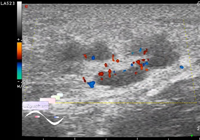
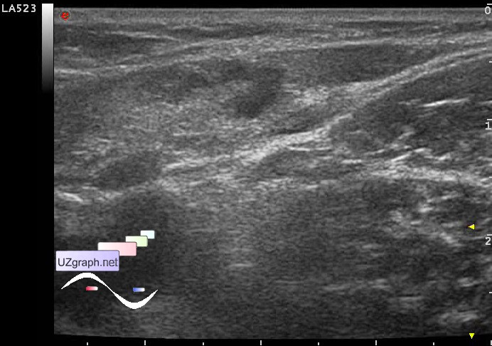
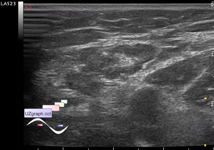
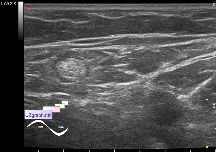
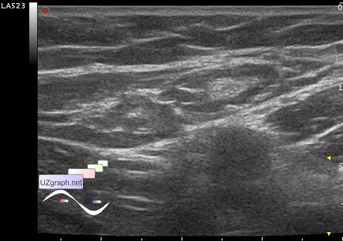
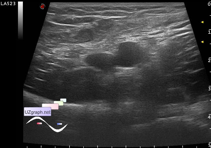
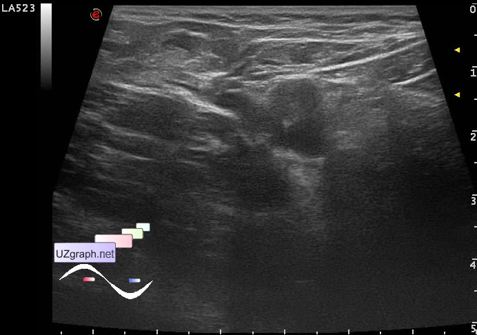
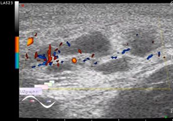
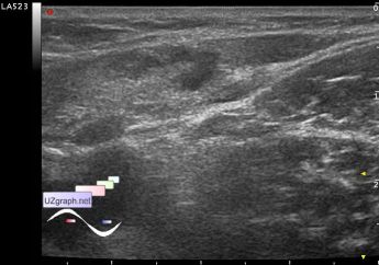
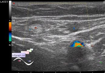
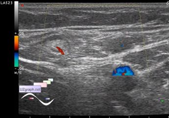
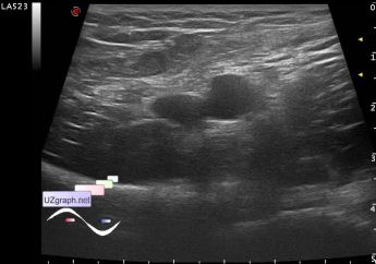
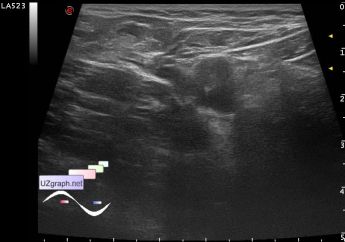
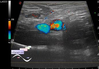
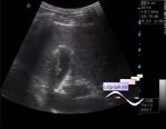
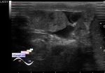
Teenager 17 years-old with complaints of pain in inguinal-femoral area occurred the day before, previously felt malaise, weakness, without temperature by his words, went to the surgeon and aimed at ultrasound of the scrotum and soft tissue of the related area. At palpation in this area is defined the consolidation of soft tissues (tuberosity) without clear boundaries, visually skin in this area not changed. On US the scrotum is unremarkable, in the inguinal-femoral area there is a mass with an unclear border, type of curved an / hypoechoic channel with rich blood supply at CFM, below another mass with similar structure but almost without blood flow, below the group of LN with a curved contour, with increased blood flow at CFM, below in the projection of proximal femoral artery located intimately a couple of LN with violation of layers ratio - the predominance of the central hyperechogenic component (lymphadenopathy of unknown origin?). In the symmetric region of contralateral leg in the proximal part of the FA is also a couple of LN with violation of layers ratio ... More ultrasound executed - abdominal cavity and kidneys are unremarkable... Recommended urgent consultations of infectionist, oncologist, surgeon. According to current information, to the child was offered a hospitalization in infectious hospital, but instead of this offer the patient with accompanying person decided to go to a some research institute, where they were told that nothing terrible and offered a treatment at home ... external link |