Log in    RU  


Main page :: Forum :: Ultrasound cases - Sonograms, cine-loops etc. ::

Bilateral popliteal cysts with legs


Tags: Soft tissues sonography, Musculoskeletal sonography, Esaote MyLab 70, Images, Video, Clinical report, Pediatric

   Posts
 
  Bilateral popliteal cysts with legs
#1

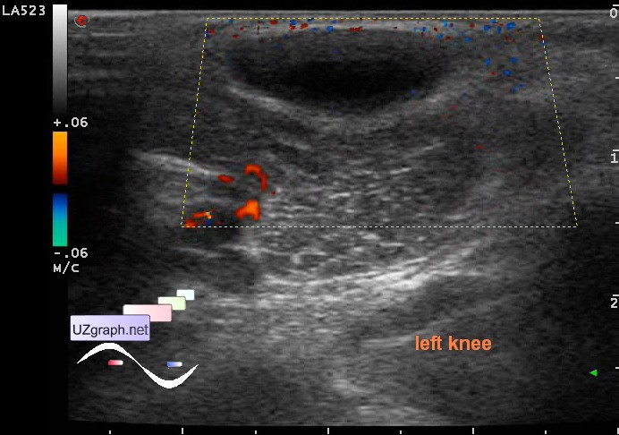
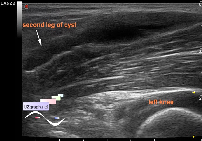
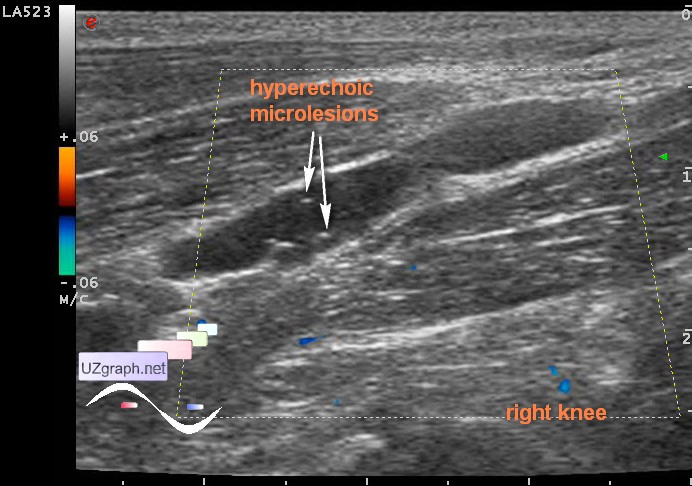
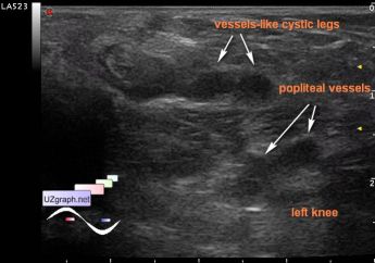
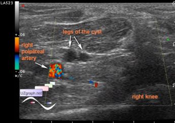
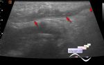
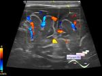
    Child 4 years-old with complaints of a painless lesion in the left popliteal area from the surgeon with suspected hygroma aimed to ultrasound of soft tissues of related area.

    Visually lesion barely visible only in the standing position, palpable ...

    At US in the left popliteal area visualized anechoic lesion, maybe a group of lesions, elongated shape - along the longitudinal axis of the body, coming from popliteal vascular bundle to the skin surface, in the initial section is represented by two parallel tubular structures(vessel-like, "legs" ), size of about 3x1,5cm, without blood flow at CFM(lymphangioma?)

    I do not know why, but decided to look for something like that in the right and found almost a twin, just a little smaller in size and with hyperechoic microlesions (microcalcifications? debris? hemorrhage in the cyst?) - bilateral lymphangioma?

    Recommended consultation of the oncologist / MRI.

    external link

:: attachments(8) ::

:: file 1 ::

:: file 2 ::

:: file 3 ::

:: file 4 ::

:: file 5 ::

:: file 6 ::

:: file 7 ::

:: file 8 ::

  20:59 17-08-2015
 
#2

    "Focal enlargement of veins, arteries, and lymphatic structures around the knee can produce a "cystic" mass. [2],[14] These conditions include aneurysms, varices, hemangiomas, and lymphangiomas.

    ...

    Lymphatic malformations are often present at birth, but may not become evident until later in life. MRI demonstrates a similar appearance to that of venous malformations; they may be macrocystic or microcystic, with the latter form predominating in the extremities"

    external link

  20:37 29-03-2018
 
#3

    Also it can be Baker's cysts...

  15:40 03-11-2019
 
#4

    According to the echo-picture in the differential diagnosis can be added a cystic adventitial disease of the popliteal artery - external link

    But in this area can be found something else -

    Peroneal intraneural ganglion cyst

    external link


 Random 
Ultrasound cases - Hemorrhagic ovarian cyst & fluid sac in uterus cervix
    

Ultrasound cases - Cholelithiasis plus
    

Ultrasound cases - Beta-thalassemia and vermiform appendix
    

Ultrasound cases - Biceps rupture
    

Ultrasound cases - Neonatal testicular torsion
    

 Dictionary 
HR

GS

CRL

LMP

RAP

TAMEAN

M-mode

GA

Aliasing doppler artifact

Anatomical M-mode


 Calculators 
Myocardial mass

Volume (ellipse formula)

Estimated fetal weight by Ultrasound

Resistive index

Type of an infant hip joint


 Similar topics 
Ultrasound cases - Penetrating injury of the femur
    

Ultrasound cases - Cephalohematoma
    

Ultrasound cases - Urachus fistula
    

Ultrasound cases - Cranial mass
    

Ultrasound cases - Pilonidal coccyx cyst
    

Ultrasound cases - Palm fistula, in search of pencil lead
    

Ultrasound cases - Postoperative abdominal wall fistula
    

Ultrasound cases - Nucks cyst
    

Ultrasound cases - Paraproctitis
    

Ultrasound cases - Submandibular area lesion