Log in    RU  


Main page :: Forum :: Ultrasound cases - Sonograms, cine-loops etc. ::

Calcific abdominal lymphadenopathy


Tags: Abdomen sonography, Soft tissues sonography, Images, Video, Clinical report, Esaote MyLab 70, Pediatric

   Posts
 
  Calcific abdominal lymphadenopathy
#1

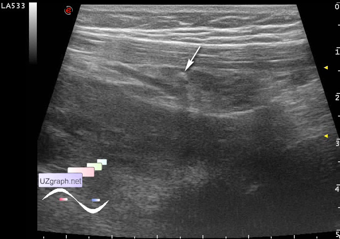
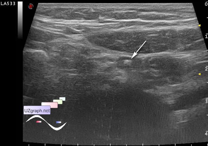
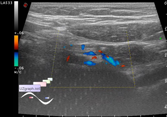
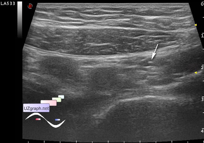
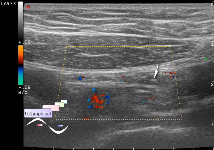
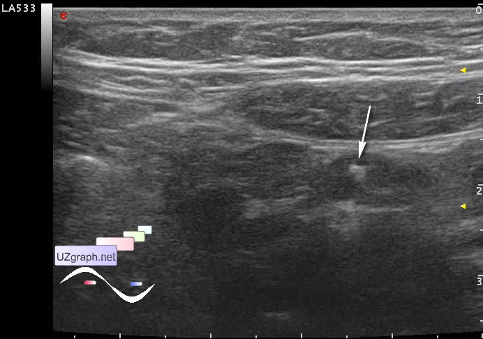
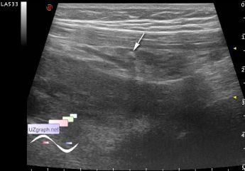
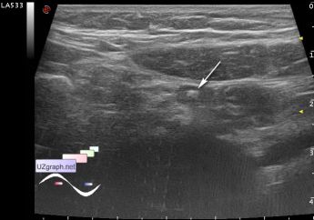
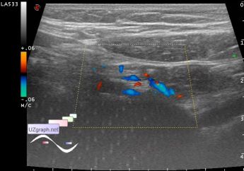
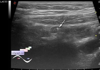
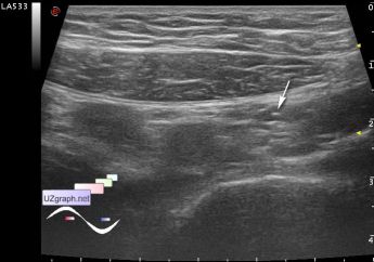
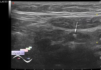
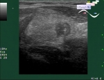
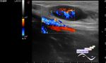
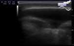
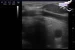
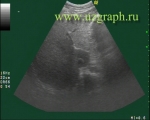
    A child 17 years old came for the screening.

    At abdominal ultrasound in mesogaster visualized more than 3 lymph nodes upto 8 mm, two of them with hyperechoic lesions (Calcific mesenteric lymphadenopathy, diff.diagnosis: tuberculosis(tbs), metastases(mts), sarcoidosis, amyloidosis, etc.)

    Part of the abdominal cavity is shielded. The wall of the stomach is thickened upto 4 mm (gastritis?).

    I asked her whether she sick some severe infections, she said that had a long time ago acute tonsillitis.

    Recommended consultation of infectionists, gastroenterologist.

    PS. Examples from a colleague's website:

    "Papillary thyroid carcinoma with lymphe node metastases with punctate calcifications..."

    external link

    "...lymph node with calcifications caused by an atypical mycobacterium"

    external link

    Diff.diagnosis:

    external link

    external link

:: attachments(8) ::

:: file 1 ::

:: file 2 ::

:: file 3 ::

:: file 4 ::

:: file 5 ::

:: file 6 ::

:: file 7 ::

:: file 8 ::


 Random 
Ultrasound cases - Tetralogy of Fallot
    

Ultrasound cases - Linea alba hernia vs lipoma part deux
    

Ultrasound cases - Starry sky sign
    

Ultrasound cases - Incomplete membrane vs abnormal chord of LA
    

Ultrasound cases - Wrist edema
    

 Dictionary 
LVESV

TAMEAN

MSD

TVUS

HC

Aliasing doppler artifact

HR

VTI

LVOT

TI-RADS


 Calculators 
Body surface area

Gestational age by Ultrasound

Myocardial mass

Type of an infant hip joint

Estimated fetal weight by Ultrasound


 Similar topics 
Ultrasound cases - Hirschsprung disease on ultrasound
    

Ultrasound cases - Intestinal Infiltrate
    

Ultrasound cases - Abdominal cryptorchidism
    

Ultrasound cases - Diffuse heterogeneous liver
    

Ultrasound cases - Postoperative abdominal wall fistula
    

Ultrasound cases - Stump appendicitis
    

Ultrasound cases - Liver scar after resection
    

Ultrasound cases - Subdiaphragmatic movable fluid collection (UFO)
    

Ultrasound cases - Lesion of the spleen or the case when it is easy to not notice
    

Ultrasound cases - I’m normal but others ...