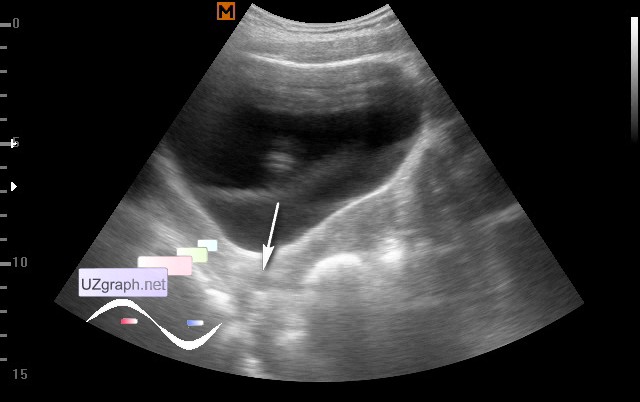
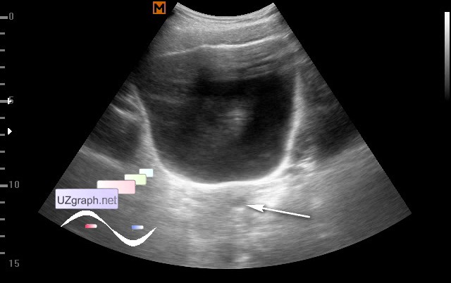
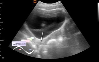
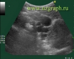
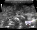
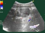
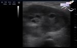
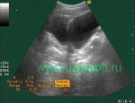
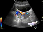
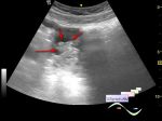
Log in    RU  


Main page :: Forum :: Ultrasound cases - Sonograms, cine-loops etc. ::

nabothian cyst


Tags: Female pelvis sonography, Medison Sonoace R7, Images, Video, Clinical report, Pediatric

   Posts
 
  nabothian cyst
#1

    The girl of 16 years, has come from the gynecologist in connection with irregular menses, earlier on ultrasound the ovarian cyst was revealed.

    At this moment, in the cervical region is visualized anechoic lesion with acoustic enhancement behind - the cervix cyst (nabothian cyst).

:: attachments(3) ::

:: file 1 ::

:: file 2 ::

:: file 3 ::


 Random 
Ultrasound cases - Thyroid lesion
    

Ultrasound cases - Thyroiditis 2
    

Ultrasound cases - 99 Luftballons
    

Ultrasound cases - Sinus venosus ASD / PAPVC
    

Ultrasound cases - Hemorrhagic ovarian cyst & fluid sac in uterus cervix
    

 Dictionary 
LVEDV

BSA

PRF

LVESV

PADP

Qp-Qs

Doppler angle

Aliasing doppler artifact

IVC

HC


 Calculators 
Myocardial mass

Body surface area

TI-RADS

Pulmonary artery pressure

Teicholz systolic cardiac function


 Similar topics 
Ultrasound cases - Right ovary changes
    

Ultrasound cases - Ovarian apoplexy 3
    

Ultrasound cases - Hemorrhagic ovarian cyst
    

Ultrasound cases - Patchy uterus
    

Ultrasound cases - Fibroid and nabothian cyst
    

Ultrasound cases - Small pelvis lesion, presumably hydrocolpos
    

Ultrasound cases - Something in the female pelvis
    

Ultrasound cases - Uterine myoma(fibroid)
    

Ultrasound cases - Giant mass in the female pelvis
    

Ultrasound cases - Gallbladder, free fluid, etc.