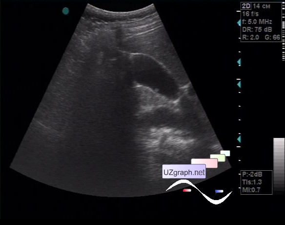
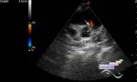
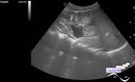
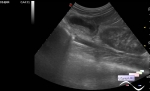
Log in    RU  


Main page :: Forum :: Ultrasound cases - Sonograms, cine-loops etc. ::

Another GB polyp


Tags: Abdomen sonography, GE Vivid 3, Images, Video, Clinical report, Adult

   Posts
 
  Another GB polyp
#1

    The female patient 24 years old, came to the abdomen ultrasound with complaints of abdominal pain.

    At ultrasonography in the projection of the posterior wall of the gallbladder is visualised lesion up to 3 mm (polyp?)

:: attachments(2) ::

:: file 1 ::

:: file 2 ::


 Random 
Ultrasound cases - Gallbladder polyps
    

Ultrasound cases - Corpus callosum dysgenesis(agenesis)


Ultrasound cases - Ileotyphlitis and/or ileocecal intussusception
    

Ultrasound cases - Accessory spleen
    

Ultrasound cases - ASD and adrenal gland lesion
    

 Dictionary 
LVPWd

Gain

PW

Freeze

AC

Color M-mode

CFM

PASP

FL

EFW


 Calculators 
Type of an infant hip joint

Pressure gradient

Myocardial mass

Gestational age by Ultrasound

Body surface area


 Similar topics 
Ultrasound cases - Portal hypertension
    

Ultrasound cases - Lesion in the projection of the adrenal gland 2
    

Ultrasound cases - Gallbladder, saw sign
    

Ultrasound cases - Ileotiphlitis and mesadenitis
    

Ultrasound cases - Cholelithiasis and hyperechoic gallbladder wall
    

Ultrasound cases - Portal vein spontaneous enhancement (mononucleosis)
    

Ultrasound cases - Hypoechoic spleen lesion
    

Ultrasound cases - Acute Appendicitis, periappendicular infiltrate
    

Ultrasound cases - Cavernous Transformation of the Portal Vein
    

Ultrasound cases - Diffuse-focal changes in the liver, thick left kidney