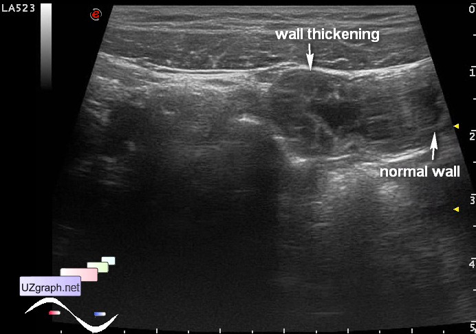
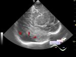
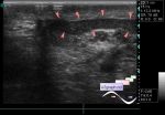
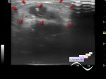
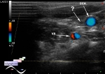
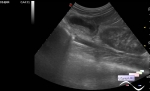
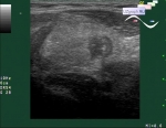
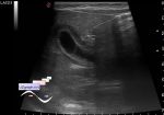
| Child 7 years old came for re-ultrasound of the abdomen and thyroid (previous examination without pathology). I asked about the cause of re-ultrasound and the cause is that child has a long-term (about a month) low-grade fever, he is already had a long course of antibiotics and several meetings with different clinicians... It is understood that an abdominal ultrasound is a very popular exam inside the medical community, the meaning of which is quite unclear for sonologists. Since in Russia most of the state healthcare is under Required Health Insurance (RHI) and such medical service, as abdominal ultrasound, in RHI not exist. Sonologist usually instead of abdominal ultrasound (who knows what is it exactly?) performed the hepatobiliary ultrasound, which is in the RHI, and includes: liver, gallbladder, bile ducts, pancreas. I.e. with this " abdomen" everything all right, now and before - a service performed for a 10 minutes (so much time in Russia now reserved for any ultrasound exam - health care reform!), with needed to clinician area - " abdomen" (translated to RHI) - job was done successful, " idiopathic" low-grade fever remained. Since I still did not struggle with idiocy by strict adherence of it, as have already begun to offer some colleagues, I decided to dig deeper, risking to receive from the next " 10 minutes patient" phrase - " Well Dr, you are working very slowly!" - now it is some kind of greeting for doctor in Russia. And I found the bilateral pyelectasis (not included in the " abdominal" ultrasound), and as it turned out after the " dig" it was a long time before, but I decided to dig a little deeper. In the right iliac area I found 2-3 mesenteric lymph nodes up to 8 mm long and a pseudokidney sign, namely the intestinal segment with irregularly thickened wall (upto 7-8 mm), presumably the ileum, with increased blood flow in the CFM ... Ie it' s likely that child has ileitis already a month because some doctors do not know what the abdominal ultrasound, while others on the contrary know exactly what it is, and sonologist hasn' t time to do more research, if he(or she) don' t want to get from the next " 10 minutes patient" phrase - " Well Dr, you are working very slowly!" .. A speed is very important, especially when catching fleas, unfortunately it is not that case, but we only have a 10 minutes ... What can we do? Can we? In 10 minutes - No way. | |