Log in    RU  


Main page :: Forum :: Ultrasound cases - Sonograms, cine-loops etc. ::

Coronary fistula


Tags: Cardiac sonography(Echocardiography), GE Logiq E, Clinical report, Pediatric

   Posts
 
  Coronary fistula
#1

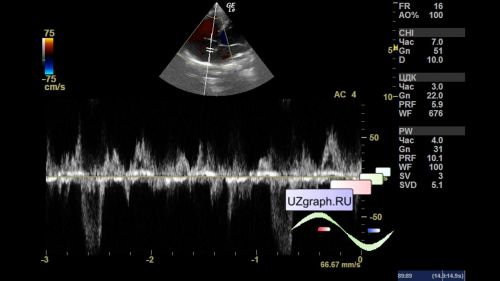
    A 6-year-old child came to the public clinic for a planned ultrasound of the heart (echocardiography) because of the murmur detected during auscultation.

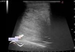
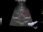
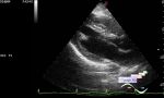
    On ultrasound with color doppler mapping: in the projection of the atrial septum, an additional flow is visualized with the direction from the septum to the right atrium, the mouth of the flow up to 3 mm, in the spectral mode in the specified projection, a high-speed flow is recorded, in the B-mode in the specified projection, a tubular structure is visualized (diff .diagnosis: coronary fistula, etc.).


 Random 
Ultrasound cases - Regional cervical lymphadenitis
    

Ultrasound cases - Dolichosigma
    

Ultrasound cases - Vortex
    

Ultrasound cases - Look at me
    

Admin room - Flash end of life

 Dictionary 
CRL

Qp-Qs

FNA

HC

LVPWd

PADP

LVOT

CFM

HR

Anatomical M-mode


 Calculators 
Myocardial mass

Gestational age by Ultrasound

Estimated fetal weight by Ultrasound

Pulmonary artery pressure

Qp:Qs


 Similar topics 
Ultrasound cases - Bicuspid aortic valve 2
    

Ultrasound cases - Mitral valve cyst
    

Ultrasound cases - Electrode in the right heart after installing of heart pacemaker
    

Ultrasound cases - Chylopericardium, lung atelectasis, cardiomegaly, ascites
    

Ultrasound cases - Asymmetric LVH, aortic regurgitation
    

Ultrasound cases - Coronary fistula from LCA?
    

Ultrasound cases - Floating IAS aneurysm
    

Ultrasound cases - Aortic Stenosis
    

Ultrasound cases - Floating Accessory Pulmonary Valve Tissue
    

Ultrasound cases - Hyperechoic linear structure in the left atrium