Log in    RU  


Main page :: Forum :: Ultrasound cases - Sonograms, cine-loops etc. ::

Bilateral single retractions of the kidney contour


Tags: Urinary tract sonography, GE Logiq E, Clinical report, Pediatric

   Posts
 
  Bilateral single retractions of the...
#1

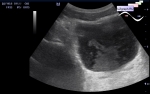
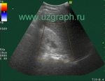
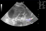
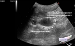
    A 5-year-old child with delayed psychomotor development and a diagnosis of tuberous sclerosis came to the public clinic for a complex ultrasound, including ultrasound of the kidneys, due to previously identified in one of the Research Institute of Pediatrics several small angiomyolipomas, the size of one of them, according to the description, is 9x2 mm, i.e. long, not round, the other was closer in size to a round shape.

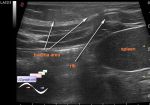
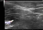
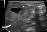
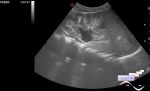
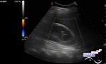
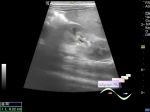
    On ultrasound, visualization is difficult, the child is constantly moving, bending, etc. is held on the couch by two attendants, in both kidneys single hyperechoic lesions are visualized according to the type of retraction of the kidney contour (folds, embryonic lobulation, etc.). These hyperechoic structures in some static views resembled rounded hyperechoic lesions in the parenchyma.


 Random 
Ultrasound cases - Extreme or good luck
    

Ultrasound cases - Nobody knows what it is
    

Ultrasound cases - Hypertrophic pyloric stenosis
    

Ultrasound cases - Patchy uterus
    

Ultrasound cases - Hydronephrosis & Ureterocele
    

 Ad 


 Dictionary 
LVOT

CFM

Anatomical M-mode

EDD

Freeze

PW

PASP

Qp-Qs

TVUS

PA


 Calculators 
Heart rate

Estimated fetal weight by Ultrasound

Myocardial mass

Teicholz formula ejection fraction

Measuring distance, area, circumference and angle on an image


 Similar topics 
Ultrasound cases - Autosomal recessive polycystic kidney disease (ARPKD)
    

Ultrasound cases - Bladder sludge
    

Ultrasound cases - Bright kidneys
    

Ultrasound cases - Ileostoma stenosis and nephropathy
    

Ultrasound cases - Renal pelvis clot
    

Ultrasound cases - Renal pelvis valve-stone
    

Ultrasound cases - Condition after treatment of a kidney cyst
    

Ultrasound cases - Hydronephrotic transformation & polypous bladder wall
    

Ultrasound cases - Pyelitis, cystitis
    

Ultrasound cases - Lesion in the projection of the adrenal gland 2
    

 Ad