Log in    RU  


Main page :: Forum :: Ultrasound cases - Sonograms, cine-loops etc. ::

Nobody knows what it is


Tags: Soft tissues sonography, Images, Video, Clinical report, Esaote MyLab 70, Pediatric

   Posts
 
  Nobody knows what it is
#1

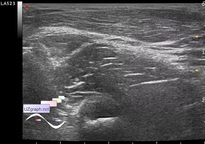
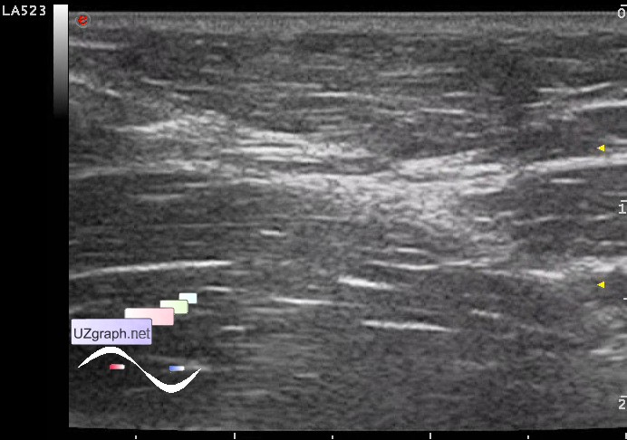
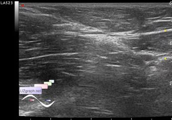
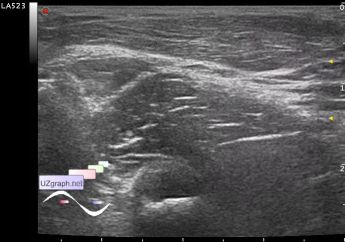
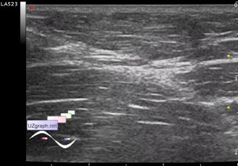
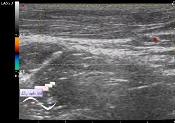
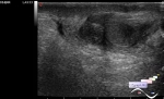
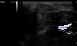
    Child 7 years-old from the surgeon with complaints about mass in inguinal-femoral area, which saw a few days ago in the shower, by the words of accompanying they had made a few US but no one knows what it is.

    Visually mass is a little above the surface of the skin, size of 3x2 cm, has the transverse orientation, skin color over mass is not changed, the medial end approaches the border of the abdominal cavity.

    On US mass has oval shape, fibrous structure, moderately moving, without blood flow at CFM (piezogenic hernia? lipoma? etc.?)

    external link

:: attachments(5) ::

:: file 1 ::

:: file 2 ::

:: file 3 ::

:: file 4 ::

:: file 5 ::

  21:46 14-01-2019
  Possible alien
#2

    Another suggestion is helminths, such as dirofilariasis.


 Random 
Ultrasound cases - Anuria. Acute renal failure
    

Ultrasound cases - Hyperechoic renal sinus with acoustic shadow
    

Ultrasound cases - Record size appendicitis
    

Ultrasound cases - Intestinal Infiltrate
    

Ultrasound cases - Indirect inguinal hernia in a girl
    

 Dictionary 
TAV

DPD

AC

LVEDV

TAMEAN

IVC

Aliasing doppler artifact

PADP

Color M-mode

EDV


 Calculators 
Pressure gradient

TI-RADS

Gestational age by Ultrasound

Pulmonary artery pressure

Thyroid volume


 Similar topics 
Ultrasound cases - Penetrating injury of the femur
    

Ultrasound cases - Cephalohematoma
    

Ultrasound cases - Urachus fistula
    

Ultrasound cases - Cranial mass
    

Ultrasound cases - Pilonidal coccyx cyst
    

Ultrasound cases - Palm fistula, in search of pencil lead
    

Ultrasound cases - Postoperative abdominal wall fistula
    

Ultrasound cases - Nucks cyst
    

Ultrasound cases - Paraproctitis
    

Ultrasound cases - Submandibular area lesion