Log in    RU  


Main page :: Forum :: Ultrasound cases - Sonograms, cine-loops etc. ::

Umbilical hernia in infant


Tags: Soft tissues sonography, Esaote MyLab 70, Clinical report, Pediatric

   Posts
 
  Umbilical hernia in infant
#1

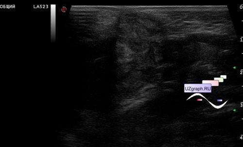
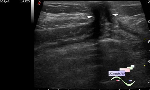
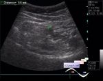
    An infant at the age of 2 months in a public clinic, sent for an ultrasound scan with a suspicion of an umbilical hernia.

    On ultrasound in the projection of the navel, an anterior abdominal wall defect up to 1 cm in size, with the contents of the abdominal cavity inside is visualized.


 Random 
Ultrasound cases - Liver cirrhosis and thickened omentum
    

Ultrasound cases - Hollow organ lesion sign, "Lost in Translation"
    

Ultrasound cases - Partial tears of the lateral meniscus
    

Ultrasound cases - Cyst but where?
    

Ultrasound cases - Newborn male mastitis
    

 Dictionary 
SF

PG

PSV

CRL

B-mode

EFW

TVUS

PA

LVESD

PRF


 Calculators 
Pressure gradient

Resistive index

Gestational age by Ultrasound

Body surface area

Pulmonary artery pressure


 Similar topics 
Ultrasound cases - Penetrating injury of the femur
    

Ultrasound cases - Cephalohematoma
    

Ultrasound cases - Urachus fistula
    

Ultrasound cases - Cranial mass
    

Ultrasound cases - Pilonidal coccyx cyst
    

Ultrasound cases - Palm fistula, in search of pencil lead
    

Ultrasound cases - Postoperative abdominal wall fistula
    

Ultrasound cases - Nucks cyst
    

Ultrasound cases - Paraproctitis
    

Ultrasound cases - Submandibular area lesion