"Imaging Findings of Chest Wall Lesions on Breast Sonography
...
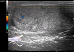
Figure 8 ... B, Schwannoma in a 29‐year‐old woman with a palpable mass in the left breast. Sonography shows a circumscribed hypoechoic mass in the subcutaneous fat layer of the chest wall.
...
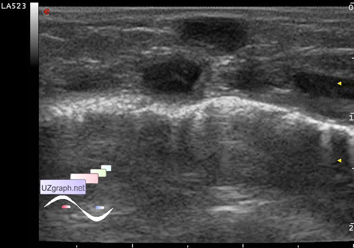
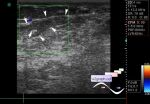
Figure 15
Lymphoma in a 60‐year‐old woman with a palpable mass below the left breast. Sonography shows a poorly defined heterogeneous hypoechoic mass with posterior acoustic enhancement in the anterior chest wall."