Log in    RU  


Main page :: Forum :: Ultrasound cases - Sonograms, cine-loops etc. ::

Rectus abdominis muscle contusion


Tags: Musculoskeletal sonography, Soft tissues sonography, Images, Video, Clinical report, Esaote MyLab 70, Pediatric

   Posts
 
  Rectus abdominis muscle contusion
#1

    8 years old girl with diffuse abdominal pain a few hours after punch from a schoolboy, aimed to abdominal ultrasound.

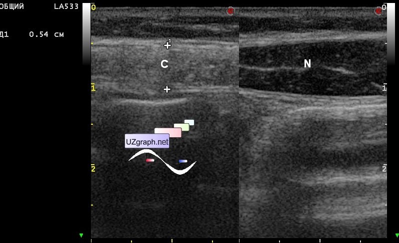
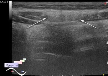
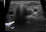
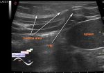
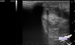
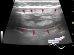
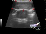
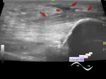
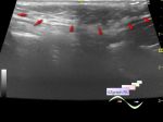
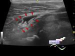
    At ultrasound internal organs of the abdomen with no complications, there is no free fluid in the abdomen also but in the upper third of the right rectus abdominis muscle is visualized wide muscle portion with increased echogenicity and a unclear border, at compression of this area with probe sharp pain revealed - presumably contusion / hematoma of the rectus abdominis muscle.

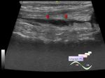
    Image legend: C - contusion, N - normal rectus abdominis muscle, scanned at the same level of other side of body.

    external link

:: attachments(3) ::

:: file 1 ::

:: file 2 ::

:: file 3 ::


 Random 
Ultrasound cases - Complication of a blood test from a vein
    

Ultrasound cases - A close friends
    

Ultrasound cases - Subhepatic lesion
    

Ultrasound cases - Good vs Evil
    

Ultrasound cases - Epididymitis
    

 Dictionary 
LMP

CFM

BSA

HR

MC

ESV

PA

TAV

CRL

ESD


 Calculators 
Body surface area

Resistive index

Gestational age by Ultrasound

Estimated fetal weight by Ultrasound

Qp:Qs


 Similar topics 
Ultrasound cases - Penetrating injury of the femur
    

Ultrasound cases - Cephalohematoma
    

Ultrasound cases - Cranial mass
    

Ultrasound cases - Recurrent shin trauma
    

Ultrasound cases - Knee joint synovitis
    

Ultrasound cases - Thickening and heterogeneity of the intercostal space soft tissues
    

Ultrasound cases - Chest wall lesion
    

Ultrasound cases - Hockey Elbow
    

Ultrasound cases - Combination of left shoulder lesions
    

Ultrasound cases - Baker's cyst and intramuscular hematoma