Log in    RU  


Main page :: Forum :: Ultrasound cases - Sonograms, cine-loops etc. ::

Hernia of the linea alba / lipoma


Tags: Soft tissues sonography, Images, Video, Clinical report, Esaote MyLab 70, Pediatric

   Posts
 
  Hernia of the linea alba / lipoma
#1

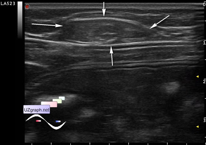
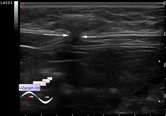
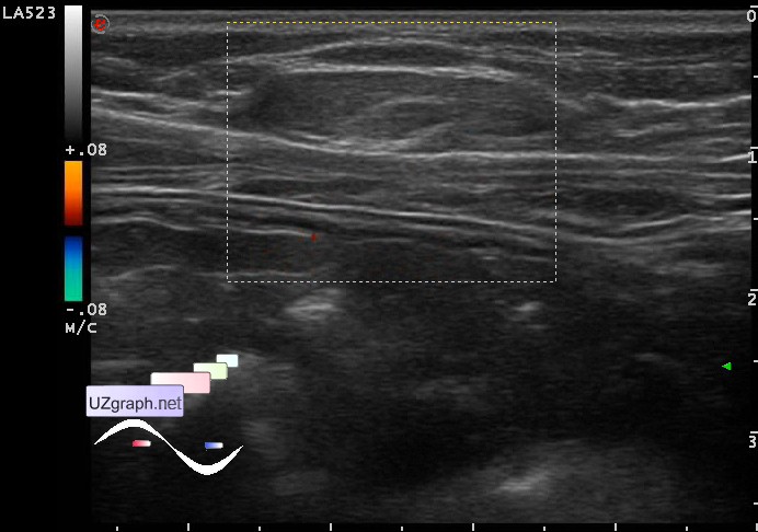
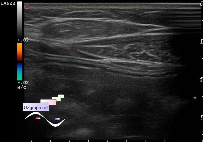
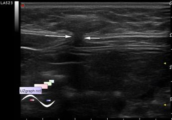
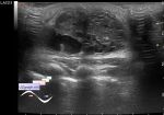
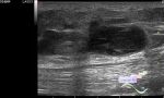
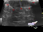
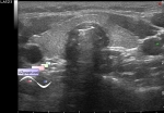
    Girl 11 y.o. with a clinical diagnosis of a linea alba hernia was sent to a the soft tissue ultrasound in public clinic after surgeon consultation.

    Visually in the center of the epigastric area there is a lesion upto 2 cm in diameter, color of the skin in the described area is not changed.

    At US visualized oval lesion about 2x0.5 cm, with similar to lipomas echo structure, at CFM there is a single area on the edge of the lesion with coloration, posterior to this lesion there is an aponeurosis defect of linea alba upto 2 mm going to the deep fatty layer. In the standing position, Valsalva maneuver - nothing changes.

    By the words of accompanying this lesion is observed from birth, in the present time solved the question of the need for surgical treatment.

    external link

:: attachments(6) ::

:: file 1 ::

:: file 2 ::

:: file 3 ::

:: file 4 ::

:: file 5 ::

:: file 6 ::

  02:00 19-05-2015
  Where is it coming from?
#2

    external link

    external link

    " 58. Linea alba and fat"

    external link

    external link

    " 1. Fat...64. Linea alba"


 Random 
Ultrasound cases - Lenticulostriate vasculopathy
    

Ultrasound cases - Cyst-like lesion in the back of the neck in infant
    

Ultrasound cases - Thyroid lesion 2
    

Scientific - Playlist Neurosonography

Ultrasound cases - Beads eggs
    

 Dictionary 
PASP

RVOT

Aliasing doppler artifact

HC

EDD

PD

DPD

ESV

Freeze

TI-RADS


 Calculators 
Type of an infant hip joint

Teicholz systolic cardiac function

TI-RADS

Volume (ellipse formula)

Pressure gradient


 Similar topics 
Ultrasound cases - Penetrating injury of the femur
    

Ultrasound cases - Cephalohematoma
    

Ultrasound cases - Urachus fistula
    

Ultrasound cases - Cranial mass
    

Ultrasound cases - Pilonidal coccyx cyst
    

Ultrasound cases - Palm fistula, in search of pencil lead
    

Ultrasound cases - Postoperative abdominal wall fistula
    

Ultrasound cases - Nucks cyst
    

Ultrasound cases - Paraproctitis
    

Ultrasound cases - Submandibular area lesion