Log in    RU  


Main page :: Forum :: Ultrasound cases - Sonograms, cine-loops etc. ::

Ovarian apoplexy 2


Tags: Female pelvis sonography, Esaote MyLab 70, Images, Video, Clinical report, Pediatric

   Posts
 
  Ovarian apoplexy 2
#1

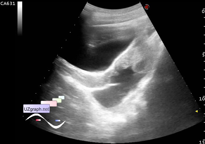
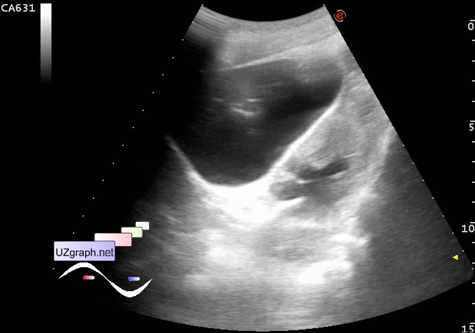
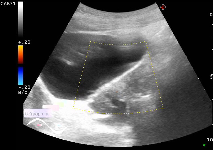
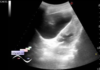
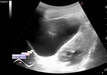
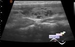
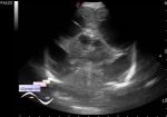
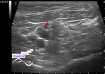
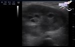
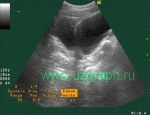
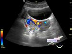
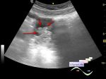
    A child 17 years old with complaints of pain in the right iliac region, which she said occurred about a week ago, has addressed to the gynecologist, it is aimed at a planned pelvis US.

    At US the M-echo to 9-10mm, at the posterior contour of the right ovary is visualized the section type of crater / discontinuation of the contour about 5-8mm (apoplexy?).

    Posterior to the uterus, more on the right, about 50 ml of free fluid.

    With suspicion of an apoplexy of the right ovary was recommended the emergency consultation with the surgeon / gynecologist. For child was called an ambulance.

    external link

    PS. Similar case - https://m.en.uzgraph.ru/forum/...

:: attachments(8) ::

:: file 1 ::

:: file 2 ::

:: file 3 ::

:: file 4 ::

:: file 5 ::

:: file 6 ::

:: file 7 ::

:: file 8 ::


 Random 
Ultrasound cases - Extreme or good luck
    

Ultrasound cases - Chest wound with barbed wire
    

Ultrasound cases - When nothing is clear
    

Ultrasound cases - Testicular torsion after surgery
    

Ultrasound cases - Nephrocalcinosis
    

 Dictionary 
GS

PSV

HR

PG

LVESD

ESD

CO

CRL

LVPWd

LN


 Calculators 
Thyroid volume

Type of an infant hip joint

Qp:Qs

Gestational age by Ultrasound

Resistive index


 Similar topics 
Ultrasound cases - Nucks cyst
    

Ultrasound cases - Hydrosalpinx (saktosalpinx)
    

Ultrasound cases - Portal vein air in the newborn
    

Ultrasound cases - Ovarian teratoma
    

Ultrasound cases - Inguinal hernia with an ovary
    

Ultrasound cases - Giant cyst of the right lateral canal
    

Ultrasound cases - Right ovary changes
    

Ultrasound cases - Ovarian apoplexy 3
    

Ultrasound cases - Hemorrhagic ovarian cyst
    

Ultrasound cases - Patchy uterus