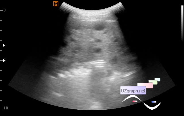
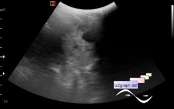
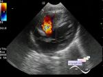
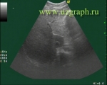
The child of 6 years, came to the surgeon, the surgeon came to me with a previous ultrasound protocol performed last month in the city polyclinic in another city where multiple anechogenic lesions in the spleen were described, the liver and kidneys were normal. As far as I understood from the words of the attendant, the child underwent a planned screening of ultrasound before entering the school in accordance with the order of the Ministry of Health of the Russian Federation. Also, in september of the previous year, the child suffered from giardiasis(lambliasis), ultrasound, as far as I understood, was not performed, the child seemed to be cured and that' s all. On the current ultrasound, everything was confirmed: spleen size 8 x 4 cm , markedly diffuse-heterogeneous structure due to multiple (10-20) anechogenous round lesions of different size, maximal up to 13 mm in diameter, at CFM without blood flow. Consultations of an infectious disease specialist and a hematologist are recommended. P.S. Perhaps lambliasis, and can echinococcosis, and others nosology no one excluded. For example, Management of splenic abscess: report on 16 cases from a single center external link Non Hodgkin' s disease with multiple hypoechoic splenic lesions external link Multiple focal lesions in liver and spleen in acute leukaemia external link Small hypoechoic lesions in liver and spleen in a patient with sarcoidosis external link Cat Scratch Disease - Splenorenal Manifestations of Bartonella henselae Infection in a Pediatric Patient external link Multiple cystic lymphangiomas of the spleen: radiologic and histopathologic findings external link external link |